Warren symposium follows legacy of geneticist giant

If we want to understand how the brain creates memories, and how genetic disorders distort the brain’s machinery, then the fragile X gene is an ideal place to start. That’s why the Stephen T. Warren Memorial Symposium, taking place November 28-29 at Emory, will be a significant event for those interested in neuroscience and genetics. Stephen T. Warren, 1953-2021 Warren, the founding chair of Emory’s Department of Human Genetics, led an international team that discovered Read more

Mutations in V-ATPase proton pump implicated in epilepsy syndrome

Why and how disrupting V-ATPase function leads to epilepsy, researchers are just starting to figure Read more

Tracing the start of COVID-19 in GA

At a time when COVID-19 appears to be receding in much of Georgia, it’s worth revisiting the start of the pandemic in early 2020. Emory virologist Anne Piantadosi and colleagues have a paper in Viral Evolution on the earliest SARS-CoV-2 genetic sequences detected in Georgia. Analyzing relationships between those virus sequences and samples from other states and countries can give us an idea about where the first COVID-19 infections in Georgia came from. We can draw Read more

Alzheimer’s Disease

Proteomics making fantastic routine

Much of basic biomedical research concerns proteins. The enzymes that keep cells running, the regulators and receptors that control what our cells do, the antibodies that defend us against invaders — all of these are proteins.

That means every day, scientists are asking questions like:

What’s happening to my favorite protein? Is there more or less of it in this sample? What other proteins work with it or stick to it?

That’s where a proteomics core facility comes in. Given a mixture of hundreds or even thousands of proteins, proteomics specialists can separate, identify and quantify them.Proteomics1smaller

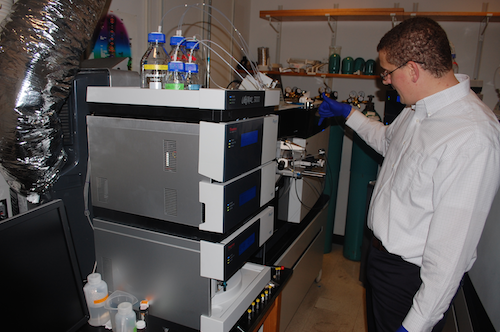
Researchers in the areas of Alzheimer’s disease, cancer metabolism, schizophrenia and vaccines all make use of Emory’s proteomics core facility. It was key to the Alzheimer’s Disease Research Center’s 2013 discovery of a new form of Alzheimer’s disease protein pathology.

Director Nick Seyfried reports that the core has acquired close to $3 million in sophisticated mass spectrometry equipment in the last few years. The Emory Integrated Proteomics Core, one of the Emory Integrated Core Facilities, is supported in part by the Winship Cancer Institute, the Atlanta Clinical and Translational Science Institute, and a recently renewed grant for ENNCF (Emory Neurosciences NINDS Core Facilities).

Protein mass spectrometry is like Wonkavision

There’s a scene in both the 1971 and 2005 film adaptations of Roald Dahl’s Charlie and the Chocolate Factory, in which a chocolate bar is separated into millions of tiny pieces and sent flying across a clean room. Protein mass spectrometry resembles the first part of this process. Read more

Posted on by Quinn Eastman in Neuro Leave a comment

NINDS director: neuroscience now largest ‘bucket of money’

On Friday, NINDS director Walter Koroshetz made an interesting remark in a lecture to Emory’s Department of Neurology. He said that in the 2016 National Institues of Health budget, neuroscience is now the largest “bucket of money,” especially with the recent boost in funding for Alzheimer’s research. That’s larger than the bucket for cancer. To be sure, biomedical research in general got a boost from Congress, with the NIH receiving its largest increase in a decade, and cancer is still a big deal!

Koroshetz explained that neuroscience research is spread out among NINDS (National Institute for Neurological Disorders and Stroke), NIMH (National Institute of Mental Health), NIDA (National Institute for Drug Abuse) and several others, while cancer research is concentrated at the National Cancer Institute. [Here’s some official category tracking that the NIH does – his breakdown checks out.]

Koroshetz highlighted a project from Dieter Jaeger and Garret Stanley that is part of the White House’s BRAIN Initiative focused on mapping brain circuits and connectivity. He also noted NINDS’s efforts in promoting translational research, since pharmaceutical companies were frustrated by repeated failures in the 1990s with difficult areas such as stroke, and the R35 mechanism for funding “outstanding investigators” for up to eight years continuously.

Posted on by Quinn Eastman in Neuro 2 Comments

The secrets of a new Alzheimer’s secretase

The title of Keqiang Ye’s recent Nature Communications paper contains a provocative name for an enzyme: delta-secretase.

Just from its name, one can tell that a secretase is involved in secreting something. In this case, that something is beta-amyloid, the toxic protein fragment that tends to accumulate in the brains of people with Alzheimer’s disease.

Aficionados of Alzheimer’s research may be familiar with other secretases. Gamma-secretase was the target of some once-promising drugs that failed in clinical trials, partly because they also inhibit Notch signaling, important for development and differentiation in several tissues. Now beta-secretase inhibitors are entering Alzheimer’s clinical trials, with similar concerns about side effects.

Many Alzheimer’s researchers have studied gamma- and beta-secretases, but a review of the literature reveals that so far, only Ye and his colleagues have used the term delta-secretase.

This enzyme previously was called AEP, for asparagine endopeptidase. AEP appears to increase activity in the brain with aging and cleaves APP (amyloid precursor protein) in a way that makes it easier for the real bad guy, beta-secretase, to produce bad beta-amyloid.*At Alzforum, Jessica Shugart describes the enzyme this way:

Like a doting mother, AEP cuts APP into bite-sized portions for toddler BACE1 [beta-secretase] to chew on, facilitating an increase in beta-amyloid production. Read more

Posted on by Quinn Eastman in Neuro Leave a comment

Do Alzheimer’s proteins share properties with prions?

If you’ve come anywhere near Alzheimer’s research, you’ve come across the “amyloid hypothesis” or “amyloid cascade hypothesis.”

This is the proposal that deposition of amyloid-beta, a major protein ingredient of the plaques that accumulate in the brains of Alzheimer’s patients, is a central event in the pathology of the disease. Lots of supporting evidence exists, but several therapies that target beta-amyloid, such as antibodies, have failed in large clinical trials.

Jucker_Walker_May_2014

Lary Walker and Matthias Jucker in Tübingen, 2014

In a recent Nature News article, Boer Deng highlights an emerging idea in the Alzheimer’s field that may partly explain why: not all forms of aggregated amyloid-beta are the same. Moreover, some “strains” of amyloid-beta may resemble spooky prions in their ability to spread within the brain, even if they can’t infect other people (important!).

Prions are the “infectious proteins” behind diseases such as bovine spongiform encephalopathy. They fold into a particular structure, aggregate and then propagate by attracting more proteins into that structure.

Lary Walker at Yerkes National Primate Research Center has been a key proponent of this provocative idea as it applies to Alzheimer’s. To conduct key experiments supporting the prion-like properties of amyloid-beta, Walker has been collaborating with Matthias Jucker in Tübingen, Germany and spent four months there on a sabbatical last year. Their paper, describing how aggregated amyloid-beta is “seeded” and spreads through the brain in mice, was recently published in Brain Pathology.
Read more

Posted on by Quinn Eastman in Neuro Leave a comment

A structure for SorLA/LR11

The importance of the SorLA or LR11 receptor in braking Alzheimer’s was originally defined here at Emory by Jim Lah and Allan Levey’s labs. Japanese researchers recently determined the structure of SorLA and published the results in Nature Structural and Molecular Biology. Their findings point toward a direct role for SorLA in binding toxic circulating beta-amyloid and transporting it to the lysosome for degradation. Hat tip to Alzforum.

Posted on by Quinn Eastman in Neuro Leave a comment

Acidity of aging leads to new Alzheimer’s drug target

Pathologist Keqiang Ye and his colleagues have been studying the functions of an enzyme called AEP, or asparagine endopeptidase, in the brain. AEP is activated by acidic conditions, such as those induced by stroke or seizure.

AEP is a protease. That means it acts as a pair of scissors, snipping pieces off other proteins. In 2008, his laboratory published a paper in Molecular Cell describing how AEP’s acid-activated snipping can unleash other enzymes that break down brain cells’ DNA.

Following a hunch that AEP might be involved in neurodegenerative diseases, Ye’s team has discovered that AEP also acts on tau, which forms neurofibrillary tangles in Alzheimer’s disease.

“We were looking for additional substrates for AEP,” Ye says. “We knew it was activated by acidosis. And we had read in the literature that the aging brain tends to be more acidic, especially in Alzheimer’s.”

The findings, published in Nature Medicine in October, point to AEP as a potential target for drugs that could slow the advance of Alzheimer’s, and may also lead to improved diagnostic tools. Read more

Posted on by Quinn Eastman in Neuro Leave a comment

Explainer: the locus coeruleus

The locus coeruleus is a part of the brain that has been getting a lot of attention recently from Emory neuroscience researchers.

The locus coeruleus is the biggest source of the neurotransmitter norepinephrine in the brain. Located deep in the brainstem, it has connections all over the brain, and is thought to be involved in arousal and attention, stress, memory, the sleep-wake cycle and balance.

Researchers interested in neurodegenerative disease want to look at the locus coeruleus because it may be one of the first structures to degenerate in diseases such as Alzheimer’s and Parkinson’s. In particular, the influential studies of German neuro-anatomist Heiko Braak highlight the locus coeruleus as a key “canary in the coal mine” indicator of neurodegeneration.

That’s why neurologist Dan Huddleston, working with biomedical imaging specialists Xiangchuan Chen and Xiaoping Hu and colleagues at Emory, has been developing a method for estimating the volume of the locus coeruleus by magnetic resonance imaging (MRI). Their procedure uses MRI tuned in such a way to detect the pigment neuromelanin (see panel), which accumulate in both the locus coeruleus and in the substantia nigra. Read more

Posted on by Quinn Eastman in Neuro Leave a comment

Beyond the usual suspects among Alzheimer’s proteins

If you’ve been paying attention to Alzheimer’s disease research, you’ve probably read a lot about beta-amyloid. It’s a toxic protein fragment that dominates the plaques that appear in the brains of people with Alzheimer’s. Many experimental therapies for Alzheimer’s target beta-amyloid, but so far, they’ve not proven effective.

That could be for several reasons. Maybe those treatments started too late to make a difference. But an increasing number of Alzheimer’s researchers are starting to reconsider the field’s emphasis on amyloid. Nature News has a feature this week explaining how the spotlight is shifting to the protein ApoE, encoded by the gene whose variation is responsible for the top genetic risk factor for Alzheimer’s.

In line with this trend, Emory’s Alzheimer’s Disease Research Center recently received a five-year, $7.2 million grant to go beyond the usual suspects like beta-amyloid. Emory will lead several universities in a project to comprehensively examine proteins altered in Alzheimer’s. You’ve heard of the Cancer Genome Atlas? Think of this as the Alzheimer’s Proteome Atlas, potentially addressing the same kind of questions about which changes are the drivers and which are the passengers.

Emory’s back-to-basics proteomics approach has already yielded some scientific fruit, uncovering changes in proteins involved in RNA splicing and processing. Also, the Nature feature also has some background on a clinical trial called TOMMORROW, which Emory’s ADRC is participating in.

Posted on by Quinn Eastman in Neuro Leave a comment

Seeing in triangles with grid cells

When processing what the eyes see, the brains of primates don’t use square grids, but instead use triangles, research from Yerkes neuroscientist Beth Buffalo’s lab suggests.

Elizabeth Buffalo, PhD

She and graduate student Nathan Killian recently published (in Nature) their description of grid cells, neurons in the entorhinal cortex that fire when the eyes focus on particular locations.

Their findings broaden our understanding of how visual information makes its way into memory. It also helps us grasp why deterioration of the entorhinal cortex, a region of the brain often affected early by Alzheimer’s disease, produces disorientation.

The Web site RedOrbit has an extended interview with Buffalo. An excerpt:

The amazing thing about grid cells is that the multiple place fields are in precise geometric relation to each other and form a tessellated array of equilateral triangles, a ‘grid’ that tiles the entire environment. A spatial autocorrelation of the grid field map produces a hexagonal structure, with 60º rotational symmetry. In 2008, grid cells were identified Gafas Ray Ban outlet in mice, in bats in 2011, and now our work has shown that grid cells are also present in the primate brain.

Please read the whole thing!

Grid cells fire at different rates depending on where the eyes are focused. Mapping that activity across the visual field produces triangular patterns.

Posted on by Quinn Eastman in Neuro 1 Comment

Tangled up with tau

Pathologist Keqiang Ye and his colleagues have identified a new potential drug target in Alzheimer’s disease. It’s called SRPK2 (serine-arginine protein kinase 2).

Keqiang Ye, PhD

Depleting this enzyme from the brain using genetic engineering tools alleviates cognitive impairment in an animal model of Alzheimer’s. The result suggests that drugs Cheap Oakleys that target this enzyme could be valuable in the treatment of Alzheimer’s, although additional studies on human brain samples are necessary to fully confirm the findings, Ye says.

The results were published Tuesday in Journal of Neuroscience. The first author is postdoctoral fellow Yi Hong.

Hong and colleagues found that SRPK2 has elevated activity in a mouse model of Alzheimer’s. It acts on tau, one of the two major toxic clumpy proteins in Alzheimer’s. (beta-amyloid is outside the cell and forms plaques, tau is inside and forms tangles). Previous research on SRPK2 indicated that it had something to do with RNA splicing, so its “entanglement” with tau is a surprise.

Posted on by Quinn Eastman in Neuro Leave a comment