Warren symposium follows legacy of geneticist giant

If we want to understand how the brain creates memories, and how genetic disorders distort the brain’s machinery, then the fragile X gene is an ideal place to start. That’s why the Stephen T. Warren Memorial Symposium, taking place November 28-29 at Emory, will be a significant event for those interested in neuroscience and genetics. Stephen T. Warren, 1953-2021 Warren, the founding chair of Emory’s Department of Human Genetics, led an international team that discovered Read more

Mutations in V-ATPase proton pump implicated in epilepsy syndrome

Why and how disrupting V-ATPase function leads to epilepsy, researchers are just starting to figure Read more

Tracing the start of COVID-19 in GA

At a time when COVID-19 appears to be receding in much of Georgia, it’s worth revisiting the start of the pandemic in early 2020. Emory virologist Anne Piantadosi and colleagues have a paper in Viral Evolution on the earliest SARS-CoV-2 genetic sequences detected in Georgia. Analyzing relationships between those virus sequences and samples from other states and countries can give us an idea about where the first COVID-19 infections in Georgia came from. We can draw Read more

Neurodegeneration accelerated by intestinal bacteria?

An influential theory about the anatomical trajectory of Parkinson’s disease is getting a microbial boost. The idea, first proposed by neuroanatomist Heiko Braak in 2003, is that pathology and neurodegeneration start in the intestines and then travel to the brain. See this article in Scientific American for background.

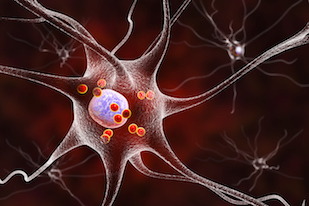
Illustration showing neurons with Lewy bodies, depicted as small red spheres, which are deposits of aggregated proteins in brain cells

Timothy Sampson, in Emory’s Department of Physiology, was first author on a recent paper in eLife, which explores the idea that prion-like proteins produced by intestinal bacteria can accelerate the aggregation of similar proteins found in our cells. The findings suggest that interventions targeting intestinal bacteria could modulate neurodegeneration.

Sampson, a former Emory graduate student who did postdoctoral work in Sarkis Mazmaniam’s lab at Caltech, says he will continue the project here. He and his colleagues were looking at the interaction between a bacterial protein called Curli – involved in adhesion + biofilms — and the aggregation-prone mammalian protein alpha-synuclein, known as a main component of the Lewy body clumps seen in Parkinson’s. The experiments were in a mouse model of Parkinson’s neurodegeneration, in which human alpha-synuclein is overproduced.

Looking ahead, Sampson says he is interested in what signals from the microbiome may trigger, accelerate or slow synuclein aggregation. He’s also looking at where in the GI tract synuclein begins to aggregate, possibly facilitated by particular cells in the intestine, and whether the observations with alpha-synuclein hold true for other proteins such as amyloid-beta in Alzheimer’s.

Posted on by Quinn Eastman in Neuro Leave a comment

Mapping the cancer genome wilderness

A huge cancer genome project has highlighted how DNA that doesn’t code for proteins is still important for keeping our cells on track.

The Pan-Cancer Analysis of Whole Genomes analyzed more than 2,600 tumors from 38 tissues, looking for causative mutations and patterns. Previous work had concentrated on the regions of the genome that code for proteins, but a significant proportion of cancer patients’ tumors don’t carry known “driver” (causative) mutations in protein-coding regions. So this project went out into what used to be called “junk DNA” or the “dark matter” of the genome.

Emory bioinformatics postdoc Matthew Reyna is the first author of one of 23 papers on the PCAWG project, published Feb. 5 in the Nature family of journals. His paper in Nature Communications looks at mutations in non-coding regions of the genome in tumors, analyzing which biological processes are affected.

Some of these were mutations in the promoters of genes encoding well-known cancer suppressors such as p53, but the project also identified new genes containing cancer-driving mutations. A promoter is the stretch of DNA that tells the cell “make RNA copies starting here”.

Reyna contributed to the project while he was at Princeton, working with Benjamin Raphael, and at Emory as well. More recently, he’s been investigating protein-protein interactions with Haian Fu, Andrey Ivanov and others as part of the Cancer Target Discovery and Development (CTD2) project.

Posted on by Quinn Eastman in Cancer Leave a comment

Stem-like CD8 T cells stay in lymph nodes/spleen

In a mouse model of chronic viral infection, there are very few virus-specific killer T cells in the blood, Emory Vaccine Center scientists report in a new paper in PNAS. This has implications for efforts to enhance cancer immunotherapy, because in both chronic viral infection and cancer, the same types of exhausted T cells accumulate.

CD8 T cells in lymphoid tissue (spleen) – from Im et al Nature (2016)

Vaccine Center director Rafi Ahmed’s lab has learned a great deal about exhausted T cells by studying the LCMV (lymphocytic choriomeningitis virus) model. In this situation, virus-specific CD8 T cells accumulate in lymph nodes and in other organs, without circulating in the blood, because they acquire a residency program, the PNAS authors write. Postdoc Sejin Im’s 2016 paper defined these “stem-like” cells – he is the first author of the new one as well.

A related phenomenon can be seen in the Kissick lab’s recent paper on immune “outposts” in kidney and other urologic tumors. The stem-like cells stay within the tumor and give rise to similar progeny. One consequence may be that treatments aimed at reactivating those cells need to get inside the tumor.

Posted on by Quinn Eastman in Immunology Leave a comment

To fight cancer, mix harmless reovirus with ‘red devil’

A recent paper in Journal of Virology mixes tried-and-true cancer-fighting tactics with the exotic. Sort of a peanut-butter-and-chocolate story, but definitely not tasty!

The tried and true is doxorubicin (Adriamycin), the notorious ‘red devil’ chemotherapy drug, which has been around for decades. On the exotic side, we have oncolytic viruses – viruses retuned to attack cancer cells more than healthy cells. This idea finally made it to FDA approval in 2015 in the form of a re-engineered herpes virus directed against melanoma.

Bernardo Mainou’s lab in the Department of Pediatrics is combining both of these approaches together. He and his team are looking to supercharge reoviruses, a mostly harmless type of virus that has been adapted into an anticancer agent. A Canadian company has brought its reovirus forward into several cancer clinical trials, but its product has not gotten to the finish line.

In the JVI paper, graduate students Roxana Rodriguez-Stewart, Jameson Berry and their colleagues infected triple-negative breast cancer cells with a variety of reoviruses, in an effort to select for those that replicate better in those cells. They also looked for drugs that enhance viral infection of those cells, and landed on doxorubicin and related drugs. Doxorubicin is part of a class of anticancer drugs that inhibit topoisomerases, enzymes that unwind DNA as part of the process of replication.

Yesterday at the GDBBS graduate research symposium, Berry gave a talk about the next step: attaching the souped-up reovirus to doxorubicin.

Three varieties of reovirus were grown together in breast cancer cells to select for efficient replication. 


Posted on by Quinn Eastman in Cancer Leave a comment

Microbiome critical for bone hormone action

Intestinal microbes are necessary for the actions of an important hormone regulating bone density, according to two papers from the Emory Microbiome Research Center. The papers represent a collaboration between Roberto Pacifici, MD and colleagues in the Department of Medicine and laboratory of Rheinallt Jones, PhD in the Department of Pediatrics.

Together, the results show how probiotics or nutritional supplementation could be used to modulate immune cell activity related to bone health. The two papers, published in Nature Communications and Journal of Clinical Investigation, are the first reports of a role for intestinal microbes in the mechanism of action of PTH (parathyroid hormone), Pacifici says.

PTH increases calcium levels in the blood and can either drive bone loss or bone formation, depending on how it is produced or administered. Continuous excessive production of PTH, or primary hyperparathyroidism, is a common endocrine cause of osteoporosis. Yet in another context, intermittent external PTH stimulates bone formation, and is an FDA-approved treatment for osteoporosis – also used off-label for fracture repair in athletes. Read more

Posted on by Quinn Eastman in Immunology Leave a comment

More NMDA but less excitotoxicity? Now possible

Emory pharmacologists have discovered a new class of potential drugs that might allow them to have their cake and eat it too — with reference to NMDA receptors, important control sites in the brain for learning and memory.

Many researchers have wanted to enhance NMDA receptor signals to treat disorders such as schizophrenia. But at the same time, they need to avoid killing neurons with “excitotoxicity”, which comes from excess calcium entering the cell. Excitotoxicity is thought to be a major mechanism of cell death in stroke and traumatic brain injury.

Usually more sensitivity to NMDA activation and excess calcium go hand in hand. In a new Nature Chemical Biology paper, pharmacologist Stephen Traynelis and colleagues have identified a group of compounds that allow them to separate those two aspects of NMDA signaling.

These compounds appear to selectively decrease how much calcium (as opposed to sodium) flows through the NMDA ion channel. Traynelis says that the discovery opens up pharmacological possibilities for NMDA receptors similar to those for other receptor classes that are prominent drug targets, such as G-protein coupled receptors and acetylcholine receptors. With such receptors, the drugs are called “biased agonists” or “biased modulators” because they shift the balance of how the ion channel responds.

For NMDA receptors, how these newly identified compounds work on a molecular level needs to be explored, and could lead to the long-standing goal of NMDA-based neuroprotection for treatment of stroke/TBI, the authors note. Postdoc Riley Perszyk is first author, with cell biologist Gary Bassell and chemists Dennis Liotta and Lanny Liebeskind as co-authors.

Traynelis discussed this research in his Hodgkin Huxley Katz Prize Lecture to the Physiology 2019 conference in Scotland in December 2019 (the part about the new class of NMDA modulators starts at about 20 minutes).

Posted on by Quinn Eastman in Neuro Leave a comment

Update on pancreatic cancer: images and clinical trial

In 2018, Winship magazine had a feature story on pancreatic cancer. Our team developed an illustration that we hoped could convey the tumors’ complex structure, which contributes to making them difficult to treat. Oncologist Bassel El-Rayes described how the tumors recruit other cells to form a protective shell.

“If you look at a tumor from the pancreas, you will see small nests of cells embedded in scar tissue,” he says. “The cancer uses this scar tissue as a shield, to its own advantage.”

With El-Rayes and fellow oncologist Walid Shaib, Greg Lesinski’s lab recently published a paper in JCI Insight. The point of the paper was to look at how chemotherapy changes immune activity in the tumor microenvironment, but we also get vivid images giving us a glimpse of those nests. It helps to view these images as large as possible, so please check them out at the journal’s site, which has no paywall.

Regions stained green are tumor-rich; red regions are immune cell-rich, and blue regions are rich in stromal cells (stellate/fibroblast cells). The goal is to get immune cells to envelop the tumors more, like in square 8.

The 2018 magazine story also laid out some of Lesinski’s and El-Rayes’ ideas.

Based on his lab’s recent success in animal models, Lesinski thinks that combining an immunotherapy drug with agents that stop IL-6 could pry open pancreatic cancers’ protective shells. In those experiments, the combination resulted in fewer stellate cells and more T cells in the tumors. Fortunately, a couple of “off-the-shelf” options, drugs approved for rheumatoid arthritis, already exist for targeting IL-6, Lesinski says.

On that theme, we noticed that a clinical trial was posted on clinicaltrials.gov in December that implements those proposals: “Siltuximab and Spartalizumab in Patients With Metastatic Pancreatic Cancer”. El-Rayes is the principal investigator, and it is not yet recruiting. Siltuximab is an antibody against IL-6 and spartalizumab is a second generation PD-1 inhibitor.

Update: The XL888 + pembrolizumab study mentioned in the article is also moving along, presented by Mehmet Akce at the Gastrointestinal Cancers Symposium.

Posted on by Quinn Eastman in Cancer Leave a comment

New animal model for elimination of latent TB

The significance of a recent Tulane/Yerkes study on eradicating latent tuberculosis in non-human primates may not be apparent at first glance. After all, it used the same antibiotic regimen (isoniazid + rifapentine) that is recommended by the CDC for human use.

But consider whether someone who was exposed to TB in childhood might still have it in their lungs somewhere. It’s difficult to know if treatments get rid of the bacteria completely.

“The antibiotic treatment we used for this study is a new, shorter regimen the CDC recommends for treating humans with latent tuberculosis, but we did not have direct evidence for whether it completely clears latent infection,” says Yerkes/Emory Vaccine Center researcher Jyothi Rengarajan, who was co-principal investigator along with Deepak Kaushal of Tulane. “Our experimental study in macaques showing almost complete sterilization of bacteria after treatment suggests this three-month regimen sterilizes humans as well.”

In an editorial in the same journal, CDC and Johns Hopkins experts call the results “dramatic” and say application of the drug regimen “could presage a major step forward in TB prevention and control.” Read more

Posted on by Quinn Eastman in Immunology Leave a comment

Transplant research: immune control via Fc receptors on T cells

Emory transplant researchers have identified a control mechanism the immune system uses to tamp down chronic inflammation. The findings provide insight into how some people were able to stop taking immunosuppressive drugs after kidney transplant.

In addition, they may be important for a full understanding of how many drugs for cancer and autoimmune disorders (therapeutic antibodies) work. The results were published on January 14 in Immunity.

In a twist, scientists have known about the molecules involved for a long time. They’re Fc receptors. Usually, we can think of them acting like oven mitts that immune cells use to grab onto antibodies. Fc receptors bind the constant (unvarying) portions of antibodies, which are the same no matter what they’re directed against.

Mandy Ford, PhD and graduate student Anna Morris

The news here is that an inhibitory variety of Fc receptor – FcγRIIB — is found on CD8+ T cells, and is a way of squeezing off T cell activity. Dogma over the past few decades held that T cells do not express Fc receptors, although evidence for them doing so went back to the 1970s.

“Our data suggest that the physiologic relevance of this pathway is to allow for control of active, highly differentiated effector T cells in the setting of chronic inflammation in order to limit immune pathology,” says senior author Mandy Ford, PhD, scientific director of Emory Transplant Center.

The co-first authors of the paper are IMP graduate student Anna Morris and surgical resident Clara Farley. They and their colleagues probed the functions of FcγRIIB on T cells in mice, and also found that increased expression of FcγRIIB correlated with freedom from rejection following withdrawal from immunosuppression in a clinical trial of kidney transplant recipients. This data came from the CTOT09 study from the Clinical Trials in Organ Transplantation Consortium. Read more

Posted on by Quinn Eastman in Immunology Leave a comment

Probing visual memory at leisure

Emory Brain Health researchers have developed a computer program that passively assesses visual memory. An infrared eye tracker monitors eye movements, while the person being tested views a series of photos.

This approach, relatively unstrenuous for those whose memory is being assessed, is an alternative for the diagnosis of mild cognitive impairment or Alzheimer’s disease. It detects degeneration of the regions of the brain that govern visual memory (entorhinal cortex/hippocampus), which are some of the earliest to deteriorate.

The approach was published in Learning and Memory last year, but bioinformatics chair Gari Clifford discussed the project at a recent talk, and we felt it deserved more attention. First author Rafi Haque is a MD/PhD student in the Neuroscience program, with neurology chair/Goizueta ADRC director Allan Levey as senior author.

Eye tracking of people with MCI and Alzheimer’s shows they spend less time checking the new or missing element in the critical region of the photo, compared with healthy controls. Adapted from Haque et al 2019.

The entire test takes around 4 minutes on a standard 24 inch monitor (a follow-up publication on an iPad version is in the pipeline). Photos are presented twice a few minutes apart, and the second time, part of the photo is missing or new – see diagram above. Read more

Posted on by Quinn Eastman in Neuro Leave a comment