Warren symposium follows legacy of geneticist giant

If we want to understand how the brain creates memories, and how genetic disorders distort the brain’s machinery, then the fragile X gene is an ideal place to start. That’s why the Stephen T. Warren Memorial Symposium, taking place November 28-29 at Emory, will be a significant event for those interested in neuroscience and genetics. Stephen T. Warren, 1953-2021 Warren, the founding chair of Emory’s Department of Human Genetics, led an international team that discovered Read more

Mutations in V-ATPase proton pump implicated in epilepsy syndrome

Why and how disrupting V-ATPase function leads to epilepsy, researchers are just starting to figure Read more

Tracing the start of COVID-19 in GA

At a time when COVID-19 appears to be receding in much of Georgia, it’s worth revisiting the start of the pandemic in early 2020. Emory virologist Anne Piantadosi and colleagues have a paper in Viral Evolution on the earliest SARS-CoV-2 genetic sequences detected in Georgia. Analyzing relationships between those virus sequences and samples from other states and countries can give us an idea about where the first COVID-19 infections in Georgia came from. We can draw Read more

Immunology

Clues to how anti-integrin antibody suppresses SIV

In October 2016, Emory and NIAID researchers published results in Science that surprised the HIV/AIDS field.

They showed that treatment with an antibody, on top of antiretroviral drugs, could lead to long-term viral suppression in SIV-infected monkeys. A similar antibody is already approved for Crohn’s disease, and a clinical trial has begun at NIAID testing the effects in people living with HIV.

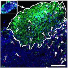
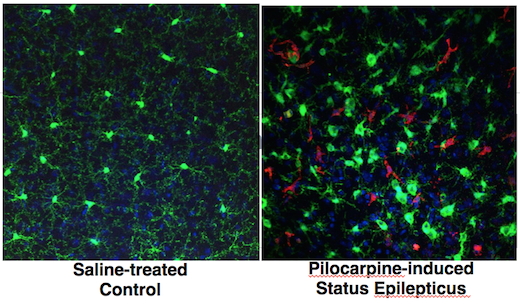
The HIV/AIDS field is still puzzling over a study led by Emory pathologist Tab Ansari.

All that was achieved even though HIV/AIDS experts are still puzzled by how the antibody works. Last week, Christina Guzzo,with NIAID director Anthony Fauci’s lab, presented new data at the Conference on Retroviruses and Opportunistic Infections in Seattle that provide some clues. But the broader issue of “what is the antibody doing?” is still open.

Let’s back up a bit. The antibody used in the Science paper targets a molecule called integrin alpha 4 beta 7, usually described as a “gut homing receptor” for CD4+ T cells, which are ravaged by HIV and SIV infection.  Study leader Aftab Ansari (right) and Fauci have both said their idea was to stop T cells from circulating into the gut, a major site of damage during acute viral infection.

Integrin alpha 4 beta 7 was also known to interact with the HIV envelope protein. Accordingly, it is possible to imagine some possibilities for what an antibody against integrin alpha 4 beta 7 could be doing: it could be driving T cells to different places in the body or affecting the T cells somehow, or it could be interfering with interactions between SIV and the cells it infects.

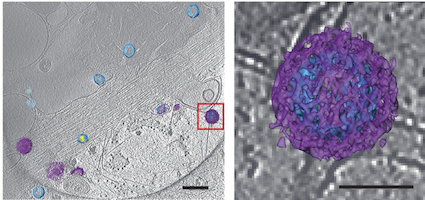
The new data from NIAID say that integrin alpha 4 beta 7 is found on the virus itself. This finding makes sense, because SIV and HIV are enveloped viruses — they steal the clothes of the cells they infect and emerge from. [Integrin alpha 4 beta 7 also appears to help the virus be more infectious in the gut, Guzzo’s presentation says.]

So a third possibility appears: the anti-alpha 4 beta 7 antibody is mopping up virus. Perhaps it’s acting like a virus-neutralizing antibody or the anti-CD4 antibody ibalizumab — CD4 is the main viral receptor on T cells. It could explain why the anti-integrin antibody’s effect is so durable; HIV/SIV can mutate to escape neutralizing antibodies directed against the viral envelope protein, but it can’t mutate the clothes it steals! Read more

Posted on by Quinn Eastman in Immunology Leave a comment

Zika virus blindfolds immune alarm cells

Important immune alarm cells — dendritic cells — are fighting Zika virus with an arm tied behind their backs, scientists from Emory Vaccine Center report.

Dendritic cells are “sentinel” cells that alert the rest of the immune system when they detect viral infection. When Zika virus infects them, it shuts down interferon signaling, one route for mustering the antiviral troops. However, another antiviral pathway called RIG-I-like receptor (RLR) signaling is left intact and could be a target for immunity-boosting therapies, the researchers say.

Mehul Suthar, PhD in the lab with graduate students Kendra Quicke and James Bowen

The findings were published on Feb. 2 in PLOS Pathogens.

Zika was known to disrupt interferon signaling, but Emory researchers have observed that it does so in ways that are distinct from other related flaviviruses, such as Dengue virus and West Nile virus. The findings give additional insight into how Zika virus is able to counter human immune defenses. Read more

Posted on by Quinn Eastman in Immunology Leave a comment

Access to HIV’s hideouts: T cells that take on their own

Police procedural television shows, such as Law + Order, have introduced many to the Internal Affairs Bureau: police officers that investigate other police officers. This group of unloved cops comes to mind in connection with the HIV/AIDS research published this week by Rama Amara’s lab at Yerkes National Primate Research Center and Emory Vaccine Center.

“Killer” antiviral T cells (red spots) can be found in germinal centers. The green areas are B cell follicles, which HIV researchers have identified as major reservoirs for the virus. Image courtesy of Rama Amara.

HIV infection is hard to get rid of for many reasons, but one is that the virus infects the cells in the immune system that act like police officers. The “helper” CD4 T cells that usually support immune responses become infected themselves. For the immune system to fight HIV effectively, the “killer” CD8 antiviral T cells would need to take on their own CD4 colleagues.

When someone is HIV-positive and is taking antiretroviral drugs, the virus is mostly suppressed but sticks around in a reservoir of inactive infected cells. Those cells hide out in germinal centers, specialized areas of lymph nodes, which most killer antiviral T cells don’t have access to. A 2015 Nature Medicine paper describes B cell follicles, which are part of germinal centers, as “sanctuaries” for persistent viral replication. (Imagine some elite police unit that has become corrupt, and uniformed cops can’t get into the places where the elite ones hang out. The analogy may be imperfect, but might help us visualize these cells.)

Amara’s lab has identified a group of antiviral T cells that do have the access code to germinal centers, a molecule called CXCR5. Knowing how to induce antiviral T cells displaying CXCR5 will be important for designing better therapeutic vaccines, as well as efforts to suppress HIV long-term, Amara says. The paper was published in PNAS this week. Read more

Posted on by Quinn Eastman in Immunology Leave a comment

Lampreys and the reverse spy problem

Call it the reverse spy problem. If you were a spy who wanted to gain access to a top secret weapons factory, your task would be to fit in. The details of your employee badge, for example, should look just right.

As described in this 2016 JCI Insight paper, Emory and University of Toronto investigators wanted to do the opposite. They were aiming to develop antibody tools for studying and manipulating plasma cells, which are the immune system’s weapons factories, where antibody production takes place. The situation is flipped when we’re talking about antibodies. Here, the goal is to stand out.

Do these guys look like good spies?

Monoclonal antibodies are classic biomedical tools (and important anticancer drugs). But it’s tricky to develop antibodies against the places where antibodies themselves are made, because of the way the immune system develops. To guard against autoimmune disease, antibodies that would react against substances in the body are often edited out.

To get around this obstacle, researchers used organisms that have very different immune systems from humans: lampreys. Emory’s Max Cooper and colleagues had already shown how lampreys have molecules — variable lymphocyte receptors or VLRs — that function like antibodies, but don’t look like them, in terms of their molecular structure.

From the paper:

We reasoned that the unique protein architecture of VLR Abs and the great evolutionary distance between lampreys and humans would allow the production of novel VLRB Abs against biomedically relevant antigens against which conventional Abs are not readily produced because of structural or tolerogenic constraints.

Senior author Goetz Ehrhardt, now at University of Toronto, used to be in Cooper’s lab, and their two labs worked together on the JCI Insight paper. Read more

Posted on by Quinn Eastman in Immunology Leave a comment

Case report on first newborn to survive Ebola

Pediatric infectious diseases specialist Anita McElroy was a co-author on a case report on the first newborn to survive Ebola infection, published recently in Journal of Infectious Diseases.

“Of all the work I’ve been privileged to be involved in over the past few years, this paper was one of the most personally satisfying,” McElroy writes.

The child described in the paper is named Nubia; she is mentioned in several news stories from 2015. She was the last known Ebola case in Guinea, one of three African countries hit hard by the virus in 2014 and 2015. Her mother died shortly after her birth.

Nubia leaves hospital in Guinea. Photo from Medecins Sans Frontieres.

Nubia was cared for at the Ebola treatment ward run by Medecins Sans Frontieres (MSF, aka Doctors without Borders) in Conakry, Guinea. She was given three experimental therapies: ZMapp antibodies, survivor white blood cell transfusion and an antiviral drug called GS-5734. It is not clear which of these interventions were critical for Nubia’s recovery, although the paper makes clear that ZMapp did not result in viral suppression all by itself.

McElroy is a go-to person for studies of dangerous viruses such as Ebola, Lassa and Zika, partly because of her affiliation with the Centers for Disease Control and Prevention’s Viral Special Pathogens Branch. She advised the MSF team on the use of the antiviral drug and other interventions.

Posted on by Quinn Eastman in Immunology Leave a comment

Double vision: seeing viruses by both light and electron microscopy

Advances in both light and electron microscopy are improving scientists’ ability to visualize viruses such as HIV, respiratory syncytial virus (RSV), measles, influenza, and Zika in their native states.

Researchers from Emory University School of Medicine and Children’s Healthcare of Atlanta developed workflows for cryo-correlative light and electron microscopy (cryo-CLEM), which were published in the January 2017 issue of Nature Protocols.

An example of the images of viruses obtainable with cryo-CLEM. Pseudotyped HIV-1 particles undergoing endocytosis. Viral membrane = light blue. Mature core = yellow. Clathrin cages = purple. From Hampton et al Nat. Protocols (2016)

Previously, many electron microscopy images of well-known viruses were obtained by studying purified virus preparations. Yet the process of purification can distort the structure of enveloped viruses, says Elizabeth R. Wright, PhD, associate professor of pediatrics at Emory University School of Medicine.

Wright and her colleagues have refined techniques for studying viruses in the context of the cells they infect. That way, they can see in detail how viruses enter and are assembled in cells, or how genetic modifications alter viral structures or processing.

“Much of what is known about how some viruses replicate in cells is really a black box at the ultrastructural level,” she says. “We see ourselves as forming bridges between light and electron microscopy, and opening up new realms of biological questions.”

Wright is director of Emory’s Robert P. Apkarian Integrated Electron Microscopy Core and a Georgia Research Alliance Distinguished Investigator. The co-first authors of the Nature Protocols paper are postdoctoral fellows Cheri Hampton, PhD. and Joshua Strauss, PhD, and graduate students Zunlong Ke and Rebecca Dillard.

The Wright lab’s work on cryo-CLEM includes collaborations with Gregory Melikyan in Emory’s Department of Pediatrics, Phil Santangelo in the Wallace H. Coulter Department of Biomedical Engineering at Georgia Tech and Emory, and Paul Spearman, now at Cincinnati Children’s.

For this technique, virus-infected or transfected cells are grown on fragile carbon-coated gold grids and then “vitrified,” meaning that they are cooled rapidly so that ice crystals do not form. Once cooled, the cells are examined by cryo-fluorescent light microscopy and cryo-electron tomography. Read more

Posted on by Quinn Eastman in Immunology Leave a comment

Four biomedical research topics to watch in 2017

HIV/AIDS

The example of the “Berlin patient,” the only person ever cured of HIV infection, has energized HIV/AIDS researchers around the world. They are exploring a variety of tactics to attack the HIV reservoir in infected people, ranging from gene editing to “kick and kill.” A host of Emory/Yerkes researchers are among those pushing this forward.

This past year, an Emory/NIAID team led by Tab Ansari showed that a gentle, antibody-based approach could suppress SIV infection in macaques for extended periods, which surprised many in the field. The human test of this approach is now underway at the National Institutes of Health.

On the preventive vaccine side, a large scale efficacy study recently begun in South Africa, the first in seven years. Geovax’s Emory-rooted technology continues to advance in clinical studies. Further back in the pipeline, Yerkes researchers are testing innovative approaches, such as Rama Amara’s milk-bacteria-based mucosal vaccine and the potent nanoparticle adjuvants developed by Bali Pulendran’s group.

Zika

Despite the World Health Organization’s declaration in November that the public health emergency is over, Zika infection is still driving brain-related birth defects in several countries. Expect to hear more about Zika epidemiology and vaccine research, including from Emory investigators, next year.

In contrast with HIV, which seems to escape from almost anything we or our immune systems throw at it, Zika is doable, scientists think. At a Vaccine Dinner Club talk in September, Harvard’s Dan Barouch made the case that Zika is a slam dunk, immunologically. Two big questions remain: does dengue get in the way? And can vaccine makers test quickly and distribute widely?

FMT for antibiotic-resistant infections

Emory physicians have been leaders in developing fecal microbiota transplant as a remedy for recurrent Clostridium dificile infection. This form of diarrhea, which can be life-threatening, sometimes arises as a result of antibiotics that wipe out the helpful bacteria that live in the intestines, paving the way for “C diff.”

Now the Emory team (Colleen Kraft/Tanvi Dhere/Aneesh Mehta/Rachel Friedman-Moraco) is testing whether FMT could prevent other antibiotic-resistant infections besides C diff. This approach will be examined in a group of patients that tends to have a lot of antibiotic exposure: kidney transplant recipients. The team’s first publication on this topic from 2014 is here. Read more

Posted on by Quinn Eastman in Immunology, Neuro Leave a comment

Threading the RSV needle: live attenuated vaccine effective in animals

Crafting a vaccine against RSV (respiratory syncytial virus) has been a minefield for 50 years, but scientists believe they have found the right balance.

A 3-D rendering of a live-attenuated respiratory syncytial virus (RSV) particle, captured in a near-to-native state by cryo-electron tomography. Surface glycoproteins (yellow) are anchored on the viral membrane (cyan), with ribonucleoprotein complexes inside (red). Image courtesy of Zunlong Ke and Elizabeth Wright.

Researchers at Emory University School of Medicine and Children’s Healthcare of Atlanta have engineered a version of RSV that is highly attenuated – weakened in its ability to cause disease – yet potent in its ability to induce protective antibodies.

The researchers examined the engineered virus using cryo-electron microscopy and cryo-electron tomography techniques, and showed that it is structurally very similar to wild type virus. When used as a vaccine, it can protect mice and cotton rats from RSV infection.

The results were published this morning in Nature Communications.

“Our paper shows that it’s possible to attenuate RSV without losing any immunogenicity,” says senior author Martin Moore, PhD, associate professor of pediatrics at Emory University School of Medicine and a Children’s Healthcare of Atlanta Research Scholar. “This is a promising live-attenuated vaccine candidate that merits further investigation clinically.”

The next steps for this vaccine are to produce a clinical grade lot and conduct a phase 1 study of safety and immunogenicity in infants, Moore says. Read more

Posted on by Quinn Eastman in Immunology Leave a comment

Insane in the membrane – inflamed in the brain

Inflammation in the brain is a feature of several neurological diseases, ranging from Parkinson’s and Alzheimer’s to epilepsy. Nick Varvel, a postdoc with Ray Dingledine’s lab at Emory, was recently presenting his research and showed some photos illustrating the phenomenon of brain inflammation in status epilepticus (prolonged life-threatening seizures).

The presentation was at a Center for Neurodegenerative Disease seminar; his research was also published in PNAS and at the 2016 Society for Neuroscience meeting.green-red-brain

Varvel was working with mice in which two different types of cells are marked by fluorescent proteins. Both of the cell types come originally from the blood and can be considered immune cells. However, one kind – marked with green — is in the brain all the time, and the red kind enters the brain only when there is an inflammatory breach of the blood brain barrier.

Both markers, CX3CR1 (green) and CCR2 (red), are chemokine receptors. Green fluorescent protein is selectively produced in microglia, which settle in the brain before birth and are thought to have important housekeeping/maintenance functions.

Monocytes, a distinct type of cell that is not usually in the brain in large numbers, are lit up red. Monocytes rush into the brain in status epilepticus, and in traumatic brain injury, hemorrhagic stroke and West Nile virus encephalitis, to name some other conditions where brain inflammation is also seen.

In the PNAS paper, Varvel and his colleagues include a cautionary note about using these mice for studying situations of more prolonged brain inflammation, such as neurodegenerative diseases: the monocytes may turn down production of the red protein over time, so it’s hard to tell if they’re still in the brain after several days.

Targeting CCR2 – good or bad? Depends on the disease model

The researchers make the case that “inhibiting brain invasion of CCR2+ monocytes could represent a viable method for alleviating several deleterious consequences of status epilepticus.” Read more

Posted on by Quinn Eastman in Immunology, Neuro Leave a comment

Antiviral success makes some immune cells stickier

As they succeed in clearing a viral infection from the body, some virus-hunting T cells begin to stick better to their target cells, researchers from Emory Vaccine Center and Georgia Tech have discovered.

The increased affinity helps the T cells kill their target cells more efficiently, but it depends both on the immune cells’ anatomic location and the phase of the infection.

The results were published this week in the journal Immunity.

Arash Grakoui, PhD

Arash Grakoui, PhD

After the peak of the infection, cells within the red pulp of the spleen or in the blood displayed a higher affinity for their targets than those within the white pulp. However, the white pulp T cells were more likely to become long-lasting memory T cells, critical for vaccines.

“These results provide a better understanding of how memory precursor populations are established and may have important implications for the development of efficacious vaccines,” the scientists write.

In the mouse model the researchers were using, the differences in affinity were only detectable a few days after the non-lethal LCMV viral infection peaks. How the differences were detected illustrates the role of serendipity in science, says senior author Arash Grakoui, PhD.

Typically, the scientists would have taken samples only at the peak (day 7 of the infection) and weeks later, when memory T cells had developed, Grakoui says. In January 2014, the weather intervened during one of these experiments. Snow disrupted transportation in the Atlanta area and prevented postdoctoral fellow Young-Jin Seo, PhD from taking samples from the infected mice until day 11, which is when the differences in affinity were apparent.

Seo and Grakoui collaborated with graduate student Prithiviraj Jothikumar and Cheng Zhu, PhD at Georgia Tech, using a technique Zhu’s laboratory has developed to measure the interactions between T cells and their target cells. Co-author Mehul Suthar, PhD performed gene expression analysis.

Read more

Posted on by Quinn Eastman in Immunology Leave a comment