If we want to understand how the brain creates memories, and how genetic disorders distort the brain’s machinery, then the fragile X gene is an ideal place to start. That’s why the Stephen T. Warren Memorial Symposium, taking place November 28-29 at Emory, will be a significant event for those interested in neuroscience and genetics.
Stephen T. Warren, 1953-2021
Warren, the founding chair of Emory’s Department of Human Genetics, led an international team that discovered Read more
At a time when COVID-19 appears to be receding in much of Georgia, it’s worth revisiting the start of the pandemic in early 2020. Emory virologist Anne Piantadosi and colleagues have a paper in Viral Evolution on the earliest SARS-CoV-2 genetic sequences detected in Georgia.
Analyzing relationships between those virus sequences and samples from other states and countries can give us an idea about where the first COVID-19 infections in Georgia came from. We can draw Read more
Why do people with cystic fibrosis (CF) have such trouble with lung infections? The conventional view is that people with CF are at greater risk for lung infections because thick, sticky mucus builds up in their lungs, allowing bacteria to thrive. CF is caused by a mutation that affects the composition of the mucus.
Rabindra Tirouvanziam, an immunologist at Emory, says a better question is: what type of cell is supposed to be fighting the bacteria?
The answer is neutrophils, one of the most abundant types of immune cells and foot soldiers against bacterial infections. When neutrophils get into the lungs in people with CF, they change behavior and shut off the expression of genes that would be important for them to combat bacteria. They stay around in the lungs, and release harmful proteins that interfere with other cells’ ability to clean up the bacteria.
Tirouvanziam’s lab has developed a culture system for studying neutrophil behavior, a model for how they act in the lungs. The system makes the neutrophils pass through a layer of lung epithelial cells. Under the influence of lung fluids obtained from CF patients, neutrophils turn what Tirouvanziam calls GRIM (Granule Release, Immunomodulatory, Metabolic). They’re feeding but not fighting: highly metabolically active, but not producing the molecules needed for bactericidal activity.
In a recent paper published in Cell Reports Medicine, researchers show that they can reverse the GRIM fate by applying alpha-amanitin, which blocks RNA transcription, and bring back bactericidal activity. This is a sledgehammer approach, because alpha-amanitin shuts down everything – it’s the toxic ingredient in destroying angel/death cap mushrooms.
Thus, alpha-amanitin would not be appropriate as a therapeutic medication. But it is a tantalizing hint of more specific approaches to come – related papers are on the way, Tirovanziam says. Reviving the anti-bacterial ability of neutrophils should be applicable regardless of the pathogen, and independent of antibiotic resistance, he adds.
“We can steer them in the right direction,” he says. “We are starting to realize that neutrophils have multiple programs and pathways – sort of like T cells. And we can show that it is being exposed to CF lung fluid that makes them go wrong – it’s not intrinsic to the neutrophils.”
The paper also says that scientists in his lab have been separating lung fluids from CF patients into fractions, in order to isolate the molecular entities responsible for steering neutrophils down the wrong path.
The first author of the Cell Reports Medicine paper was former graduate student Camila Margaroli, currently a postdoc at UAB. Tirouvanziam’s lab is part of Emory’s Department of Pediatrics and the Emory-Children’s Healthcare Center for Cystic Fibrosis and Airways Disease Research.
How long does COVID-19 vaccine-generated immunity last? New laboratory results provide a partial answer to that question.
Antibodies generated by a currently available COVID-19 vaccine declined over time, but remained at high levels in 33 study participants 6 months after vaccination, according to data published Tuesday in the New England Journal of Medicine.
The results could begin to inform public health decisions about COVID-19 booster vaccinations and how frequently people should receive them. In older study participants, antiviral antibody activity tended to decay more rapidly than in those aged 18-55.
From Doria-Rose et al (2021). Note that neutralizing antibody activity was (on average) higher at day 209 than on day 29, when the second vaccine dose was administered. It takes two weeks for the immune system to kick into high gear after the second shot.
Emory Vaccine Center’s Mehul Suthar, co-lead author of the brief report, said that the “correlates of protection” are not yet known from COVID-19 vaccine studies – that is, what levels of antiviral antibodies are needed to fend off infection. Other forms of immunity, such as T cells, could be contributing to antiviral protection as well.
He cautioned that the decay in antibody activity over time – not surprising in itself – may combine with increased prevalence of emerging SARS-CoV-2 variants that may allow viruses to escape the immune system’s pressure.
“Still, these are encouraging results,” Suthar says. “We are seeing good antibody activity, measured three different ways, six months after vaccination. There are differences between age groups, which are consistent with what we know from other studies.”
The findings come from analysis of samples from the Moderna mRNA-1273 phase I clinical trial, which began last year. Reports of clinical outcomes from Pfizer/BioNTech also indicate that their vaccine remains effective after six months.
Researchers interested in Alzheimer’s and other neurodegenerative diseases are focusing their attention on microglia, cells that are part of the immune system in the brain.
Author Donna Jackson Nakazawa titled her recent book on microglia “The Angel and the Assassin,” based on the cells’ dual nature; they can be benign or malevolent, either supporting neuronal health or driving harmful inflammation. Microglia resemble macrophages in their dual nature, but microglia are renewed within the brain, unlike macrophages, which are white blood cells that infiltrate into the brain from outside.
At Emory, neurologist Srikant Rangaraju’s lab recently published a paper in PNAS on a promising drug target on microglia: Kv1.3 potassium channels. Overall, the results strengthen the case for targeting Kv1.3 potassium channels as a therapeutic approach for Alzheimer’s.
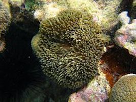
Kv1.3 potassium channels have also been investigated as potential therapeutic targets in autoimmune disorders, since they are expressed on T cells as well as microglia. The peptide dalazatide, based on a toxin from the venom of the Caribbean sea anemone Stichodactyla helianthus, is being developed by the Ohio-based startup TEKv Therapeutics. The original venom peptide needed to be modified to make it more selective toward the right potassium channels – more about that here.
Kv1.3 potassium channels are potential therapeutic targets in autoimmune disorders and Alzheimer’s — blockable with peptides based on venom of the sea anemone Stichodactyla helianthus
It appears that Kv1.3 levels on microglia increase in response to exposure to amyloid-beta, the toxic protein fragment that accumulates in the brain in Alzheimer’s, and Kv1.3 may be an indicator that microglia are turning to the malevolent side.
In the Emory paper, researchers showed that Kv1.3 potassium channels are present on a subset of microglia isolated from Alzheimer’s patients’ brains. They also used bone marrow transplant experiments to show that the immune cells in mouse brain that express Kv1.3 channels are microglia (internal brain origin), not macrophages (transplantable w/ bone marrow).
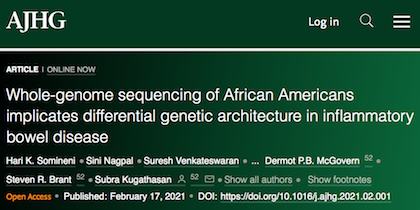
In African Americans, the genetic risk landscape for inflammatory bowel disease (IBD) is very different from that of people with European ancestry, according to results of the first whole-genome study of IBD in African Americans. The authors say that future clinical research on IBD needs to take ancestry into account.
Findings of the multi-center study, which analyzed the whole genomes of more than 1,700 affected individuals with Crohn’s disease and ulcerative colitis and more than 1,600 controls, were published on February 17 in the American Journal of Human Genetics.
As part of their analysis, the researchers developed an algorithm that corrects for ancestry when calculating an IBD polygenic risk score. Polygenic risk scores are tools for calculating gene-based risk for a disease, which are used for IBD as well as other complex conditions such as coronary artery disease.
“Even though the disease destination looks the same, the populations look very different, in terms of what specific genes contribute to risk for IBD,” says lead author Subra Kugathasan, MD. “It shows that you can’t develop a polygenic risk score based on one population and apply it to another.”
Kugathasan is scientific director of the pediatric IBD program and director of the Children’s Center for Transplantation and Immune-mediated Disorders at Children’s Healthcare of Atlanta, as well as Marcus professor of pediatrics and human genetics at Emory University School of Medicine.
The first author of the paper is geneticist Hari Somineni, PhD, who earned his doctorate working with Kugathasan at Emory, and is now working at Goldfinch Bio in Massachusetts.
The primary sites to recruit study participants were Emory, Cedars-Sinai and Rutgers, along with Johns Hopkins and Washington University at Saint Louis. Along with Kugathasan, the co-senior authors and co-organizers of the study were Steven Brant, MD from Rutgers and Dermot McGovern, MD, PhD from Cedars-Sinai.
“One of our goals in treating IBD is to move toward a more personalized approach,” says McGovern, the Joshua L. and Lisa Z. Greer Chair in Inflammatory Bowel Disease Genetics at Cedars-Sinai. “Deciphering the genetic architecture is an important part of this effort. Studies such as this one are vital to ensure that diverse populations, including African-Americans, benefit from the tremendous advances promised by genomic medicine.”
As part of an effort to strengthen genomic surveillance for emerging strains of SARS-CoV-2, the Centers for Disease Control and Prevention (CDC) has awarded a contract to Emory University researchers to characterize viral variants circulating in Georgia.
The two-year contract is part of the SPHERES (SARS-CoV-2 Sequencing for Public Health Emergency Response, Epidemiology and Surveillance) initiative, with roughly $620,000 in total costs. The principal investigator is Anne Piantadosi, MD, PhD, assistant professor of pathology and laboratory medicine, with co-investigator Mehul Suthar, PhD, assistant professor of pediatrics (infectious diseases).
Both Piantadosi and Suthar are affiliated with Emory University School of Medicine and Emory Vaccine Center. Additional Emory partners include assistant professor of medicine Ahmed Babiker, MBBS, assistant professor of medicine Jesse Waggoner, MD and assistant professor of biology Katia Koelle, PhD.
“We are analyzing SARS-CoV-2 genomes from patients in Georgia to understand the timing and source of virus introduction into our community,” Piantadosi says. “We want to know whether there have been population-level changes in the rates of viral spread, and whether there are associations between viral genotype, viral phenotype in vitro, and clinical phenotype or clinical outcome.”
The Emory MVA COVID-19 vaccine induces protective immunity with the platform of modified vaccinia Ankara (MVA), a harmless version of a poxvirus that is well-known for its use in HIV/AIDS vaccines. Like the Moderna and Pfizer COVID-19 vaccines, the Emory MVA COVID-19 vaccine induces strong neutralizing antibodies, which support the immune system’s ability to fight infections. The Emory MVA COVID-19 vaccine also induces killer CD8 T cells, providing a multi-pronged approach to halting SARS-CoV-2.
In addition, the Emory researchers say the vaccine is easily adaptable to address disease variants and can be used in combination with existing vaccines to improve their ability to combat variants and has the potential to be equally effective with a single dose.
Lead researcher Rama Amara, PhD, built the Emory MVA COVID-19 vaccine based on his more than 20 years of experience working with MVA and animal models to develop an HIV/AIDS vaccine. He and his Yerkes-based research team tested two MVA SARS-CoV-2 vaccines in mice. One of them, MVA/S, used the complete spike protein of coronavirus to induce strong neutralizing antibodies and a strong killer CD8 T cell response against SARS-CoV-2.
“Generating neutralizing antibodies is an important component of a successful COVID-19 vaccine because the antibodies can block the virus from entering the body’s cells,” says Amara, Charles Howard Candler professor of microbiology and immunology at Emory University School of Medicine and a researcher in Yerkes’ Division of Microbiology and Immunology and Emory Vaccine Center. “It’s as important to activate CD8 T cells that can clear infected cells, so this allows us to approach halting the virus two ways simultaneously. The CD8 T cells also provide ongoing value because they are key to working against other variants of the virus, especially if antibodies fail.”
Amidst the tumult in the nation’s capital, a quieter reckoning was taking place this week for the Moderna COVID-19 vaccine clinical trial. Lab Land has been hearing from Emory-affiliated study participants that they’re finding out whether they received active vaccine or placebo.
For example, Emory and Grady physician Kimberly Manning, who had written about her participation in the Moderna study in a Lancet essay, posted on Twitter Tuesday. She discovered she had received placebo, and then was offered active vaccine.
After Moderna reported strong efficacy and an Emergency Use Authorization came from the FDA, this was going to happen at some point – the question was when and how. At the advisory panel hearing in December, there was some tension over whether to remove the blind immediately, as this STAT article describes:
“Companies have said that they feel an ethical obligation to deliver vaccine to placebo recipients; the FDA and experts at its advisory panel have debated whether this obligation even exists. Instead, they argue, offering vaccine to volunteers receiving placebo limits the quality of the data about the vaccine’s long-term efficacy and side effects.”
A plan to keep participants in the study under a blinded crossover design was floated, but not implemented. Some participants have said they sensed from the start, based on temporary unpleasant side effects, whether they had received active vaccine or placebo.
For COVID-19, many researchers around the world have tried to repurpose drugs for other indications, often unsuccessfully. New clinical trial results show that baricitinib, developed by Eli Lilly and approved for rheumatoid arthritis, can speed recovery and may reduce mortality in some groups of hospitalized COVID-19 patients.
How did this study, sponsored by the National Institute of Allergy and Infectious Diseases, come together? In part, through decade-long groundwork laid by investigators at Emory, and their collaborations with others.
For several years, drug hunter and virologist Raymond Schinazi and his team had been investigating a class of medications called JAK inhibitors, as an option for tamping down chronic inflammation in HIV infection. Schinazi was one of the first at Emory to investigate the use of anti-inflammatory agents for herpesviruses and HIV in combination with antiviral drugs. He believed that these viruses “hit and run,” leaving behind inflammation, even if they later go into hiding and seem to disappear.
In Schinazi’s lab, Christina Gavegnano had shown that JAK inhibitors had both anti-inflammatory and antiviral properties in the context of HIV — a project she started as a graduate student in 2010. JAK refers to Janus kinases, which regulate inflammatory signals in immune cells.
“Our team was working on this for 10 years for HIV,” Gavegnano says. “There was a huge amount of data that we garnered, showing how this drug class works on chronic inflammation and why.”
In the race to halt the COVID-19 pandemic, researchers at Yerkes National Primate Research Center of Emory University share two important findings from their latest peer-reviewed, published study in Cell.
Rhesus monkeys are a valid animal model for COVID-19 studies because the way they experience and respond to the virus has comparable similarities to the way the virus affects humans, the researchers say. And baricitinib, an anti-inflammatory medication that is FDA-approved for rheumatoid arthritis, is remarkably effective in reducing the lung inflammation COVID-19 causes when the medication is started early after infection.
The study results have immediate and important implications for treating patients with COVID-19. Baricitinib will be compared against the steroid dexamethasone in a NIAID-sponsored clinical trial called ACTT-4 (Adaptive COVID-19 Treatment Trial), which started in November.
Mirko Paiardini, PhD, a researcher in Yerkes’ Microbiology and Immunology division, and his team selected rhesus macaques as the animal model because they expected the monkeys would mimic the disease course in humans, including the virus traveling to the upper and lower airways, and causing high levels of inflammation in the lungs. The team randomized eight rhesus macaques into two groups – a control and a treatment group; the animals in the treatment group received baricitinib.
“Our results showed the medication reduced inflammation, decreased inflammatory cells in the lungs and, ultimately, limited the virus’ internal path of destruction,” Paiardini says. “Remarkably, the animals we treated with baricitinib rapidly suppressed the processes responsible for inducing lung inflammation, thus elevating baricitinib for consideration as a frontline treatment for COVID-19 and providing insights on the way the drug works and its effectiveness.”
The FDA recently granted baricitinib emergency use authorization in combination with remdesivir based on the results of the ACTT-2 findings. “Our study was under way concurrently and, now, solidifies the importance of baricitinib in treating COVID-19,” Paiardini adds.
Co-senior author Raymond Schinazi, PhD, DSc, inventor of the most commonly used HIV/AIDS drugs to prevent progression of the disease and death, says: “Our study shows the mechanisms of action are consistent across studies with monkeys and clinical trials with humans. This means the nonhuman primate model can provide enough therapeutic insights to properly test anti-inflammatory and other COVID-19 therapies for safety and effectiveness.”
Schinazi is the Frances Winship Walters Professor of Pediatrics at Emory University School of Medicine and is affiliated with Yerkes.
“Ray and his group have been investigating the potential of anti-inflammatory drugs, such as baricitinib, for years in the context of another infection, HIV, in which inflammation is a key cause of sickness and death,” Paiardini says. “Our laboratories have collaborated for years to test therapeutics in the nonhuman primate model of HIV infection, thus placing us in a unique position when COVID-19 hit the U.S. to focus our combined expertise and efforts to halt the virus. It took only a phone call between the two of us to switch gears, begin work to create a reliable and robust monkey model of COVID-19 at Yerkes and test the potential of drugs to block inflammation.”
Tim Hoang, first author and Emory doctoral student in the Immunology and Molecular Pathogenesis Program, says: “It was exciting to be at the forefront of the response to COVID-19 and to be part of this research team that involved collaboration from Yerkes and Emory infectious disease experts, geneticists, chemists, pathologists and veterinarians.”
Co-first author and Emory postdoctoral fellow Maria Pino, PhD, emphasizes: “We knew Yerkes was uniquely suited to conduct this study because of the research and veterinary expertise, specialized facilities and animal colony, and our team’s commitment to providing better treatment options for people who have COVID-19.”
The research team plans to conduct further studies to better understand the inflammation the virus causes and to develop more targeted approached to mitigate the damage COVID-19 leaves behind.
Steven Bosinger, PhD, co-senior author, and his research team conducted the genomic analyses that helped unravel the process by which baricitinib reduces inflammation. “One of the most exciting aspects of this project was the speed genomics brought to the collaborative research,” says Bosinger. “Eight months ago, we began using genomics to accelerate the drug screening process in order to identify treatable, molecular signatures of disease between humans and model organisms, such as the monkeys in this study, In addition to determining the effectiveness of baricitinib, this study highlights Emory researchers’ commitment to improving human health and, in this case, saving human lives.”
Bosinger is assistant professor, Department of Pathology & Laboratory Medicine, Emory School of Medicine (SOM) and Emory Vaccine Center (EVC); director, Yerkes Nonhuman Primate Genomics Core and a researcher in Yerkes’ Division of Microbiology and Immunology.
Some of the others on the Emory research team include: Arun Boddapati (co-first author), Elise Viox, Thomas Vanderford, PhD, Rebecca Levit, MD, Rafick Sékaly, PhD, Susan Ribeiro, PhD, Guido Silvestri, MD, Anne Piantadosi, MD, PhD, Sanjeev Gumber, BVSc, MVSc, PhD, DACVP, Sherrie Jean, DVM, DACLAM, and Jenny Wood, DVM, DACLAM. Jacob Estes, PhD, at Oregon Health & Science University also collaborated.
Paiardini says, “So many colleagues had a key role in this study. First authors Tim and Maria as well as Yerkes veterinary and animal care personnel who worked non-stop for months on this project. This truly has been a collaborative effort at Emory University to help improve lives worldwide.”
This study was funded by the National Institutes of Health, Emory University’s COVID-19 Molecules and Pathogens to Populations and Pandemics Initiative Seed Grant, Yerkes’ base grant, which included support for the center’s Coronavirus Pilot Research Project grants, and Fast Grants.
Grant amounts (direct + indirect) are:
NIH R37AI141258, $836,452/yr (2018-23)
NIH R01AI116379, $783,714/yr (2015-20 + 2021 NCE)
NIH P51 OD011132, $10,540,602/yr (2016-20)
U24 AI120134 $681,214/yr (2020-2025)
S10OD026799 $985,030/yr (2019-2020)
Emory University COVID-19 Molecules and Pathogens to Populations and Pandemics Initiative Seed Grant, $150,000/1 yr
Fast Grants #2144, $100,000/1 yr
Note: Only a portion of the NIH grant funding was applied to the study reported in this news release.
On Thursday and Friday, Emory researchers participated in an online NIAID workshop about “post-acute sequelae” of COVID-19, which includes people with long COVID.
Long COVID has some similarities to post-viral ME/CFS (myalgic encephalomyelitis/ chronic fatigue syndrome), which has a history of being dismissed or minimized by mainstream medicine. In contrast, the workshop reflected how seriously NIAID and researchers around the world are taking long COVID.
Post-acute is a confusing term, because it includes both people who were hospitalized with COVID-19, sometimes spending weeks on a ventilator or in an intensive care unit, as well as members of the long COVID group, who often were not hospitalized and did not seem to have a severe infection to begin with.
COVID-19 infection can leave behind lung or cardiac damage that could explain why someone would have fatigue and shortness of breath. But there are also signs that viral infection can perturb other systems of the body, leading to symptoms such as “brain fog” (cognitive/memory problems), persistent pain and/or loss of smell and taste.
One goal for the workshop was to have experts discuss how to design future studies, or how to take advantage of existing studies to gain insights. A major clue on what to look for comes from Emory immunologist Ignacio Sanz, who spoke at the conference.
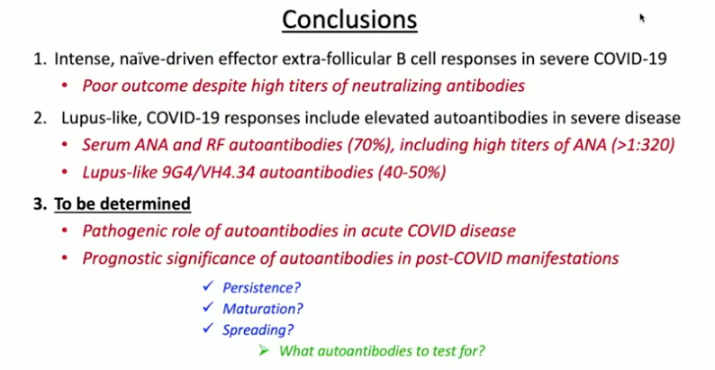
Sanz’s research has shown similarities between immune activation in people hospitalized at Emory with severe COVID-19 and in people with the autoimmune disease lupus. In lupus, the checks and balances constraining the immune system break down. A characteristic element of lupus are autoantibodies: antibodies that recognize parts of the body itself. Their presence in COVID-19 may be an explanation for the fatigue, joint pain and other persistent symptoms experienced by some people after their acute infections have passed.
Part of Ignacio Sanz’s talk at the NIAID conference on post-acute sequelae of COVID-19
For details on Sanz’s research, please see our write-up from October, their Nature Immunology paper, and first author Matthew Woodruff’s explainer. The Nature Immunology paper’s results didn’t include measurement of autoantibodies, but a more recent follow-up did (medRxiv preprint). More than half of the 52 COVID-19 patients tested positive for autoantibodies at levels comparable to those in lupus. In those with the highest amounts of the inflammatory marker CRP, the proportion was greater.
“It could be that severe viral illness routinely results in the production of autoantibodies with little consequence; this could just be the first time we’re seeing it,” Woodruff writes in a second explainer. “We also don’t know how long the autoantibodies last. Our data suggest that they are relatively stable over a few weeks. But, we need follow-up studies to understand if they are persisting routinely beyond infection recovery.”
Sanz’s group was looking at patients’ immune systems when both infection and inflammation were at their peaks. They don’t yet know whether autoantibodies persist for weeks or months after someone leaves the hospital. In addition, this result doesn’t say what is happening in the long COVID group, many of whom were not hospitalized.
It makes sense that multiple mechanisms could explain post-COVID impairments, including persistent inflammation, damage to blood vessels or various organs, and blood clots/mini-strokes.
Anthony Komaroff from Harvard, who chaired a breakout group on neurology/psychiatry, said the consensus was that so far, direct evidence of viral infection in the brain is thin. Komaroff said that neuro/psych effects are more likely to come from the immune response to the virus.
There were breakout groups for different areas of investigation, such as cardiovascular, and gastrointestinal. Emory Vaccine Center director Rafi Ahmed co-chaired a session for immunologists and rheumatologists, together with Fred Hutch’s Julie McElrath.
Emory’s Carlos del Rio, who recently summarized long COVID for JAMA, spoke about racial and ethnic disparities in COVID-19’s impact and said he expected similar inequities to appear with long COVID.
Reports from the breakout groups Friday emphasized the need to design prospective studies, which would include people before they became sick and take baseline samples. Some suggestions came for taking advantage of samples from the placebo groups in recent COVID-19 vaccine studies.