Quinn Eastman

BioArt: amyloid in the heart

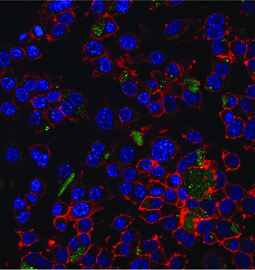
What Abstract Expressionist artist painted this? Jackson Pollock?LewisW2013

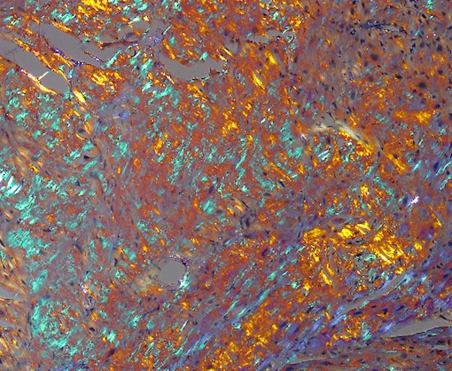
Actually, the photo depicts amyloid plaques, a frequent topic in the context of Alzheimer’s disease. Pathologist William Lewis‘ photo reminds us that amyloid can also appear in the heart.

Amyloidosis of the heart is a set of complex diseases caused by the accumulation of cellular proteins that form an amyloid plaque. Although http://www.oakleyonorder.com/ amyloidosis was described more than 100 years ago, the causative proteins were not identified until recent chemical analyses were conducted. This image shows an amyloid plaque stained with Congo red stain and viewed through a polarized lens. The optical properties of the amyloid-forming protein cause it to appear green, while other matrix materials within the plaque appear as orange and blue.

The photo, which was one of the winners of the FASEB (Federation of American Societies for Experimental Biology) 2013 BioArt competition, was featured on NIH director Francis Collins’ blog this week.

Lewis, who studies the effects of antiretroviral drugs on the cardiovascular system in his laboratory, reports that he came across the amyloid tissue sample as part of his duties as director of cardiovascular pathology: “It was beautiful.”

Posted on by Quinn Eastman in Heart Leave a comment

Fragile but potent: RNA delivered by nanoparticle

An intriguing image for November comes from biomedical engineer Mike Davis’ lab, courtesy of BME graduate student Inthirai Somasuntharam.

Each year, thousands of children undergo surgery for congenital heart defects. A child’s heart is more sensitive to injury caused by interrupting blood flow during surgery, and excess reactive oxygen species are a key source of this damage.

Macrophages with blue nuclei and red cytoskeletons, being treated with green nano particles. The particles carry RNA that shut off reactive oxygen species production.

Macrophages with blue nuclei and red cytoskeletons, being treated with green nano particles. The particles carry RNA that shut off reactive oxygen species production.

Davis and his colleagues are able to shut off cheap oakley reactive oxygen species at the source by targeting the NOX (NADPH oxidase*) enzymes that produce them. This photo, from a 2013 Biomaterials paper, shows green fluorescent nanoparticles carrying small interfering RNA. The RNA precisely shuts down one particular gene encoding a NOX enzyme. Eventually, similar nanoparticles may shield the heart from damage during pediatric heart surgery.

In the paper, Somasuntharam used particles made of a slowly dissolving polymer called polyketals. The particles delivered fragile but potent RNA molecules into macrophages, inflammatory cells that swarm into cardiac tissue after a heart attack. Davis and Georgia Tech colleague Niren Murthy previously harnessed this polymer to deliver drugs that can be toxic to the rest of the body.

The polyketal particles are especially well-suited for delivering a payload to macrophages, since those types of cells (as the name implies) are big eaters. Davis reports his lab has been working on customizing the particles so they can deliver RNA molecules into cardiac muscle cells as well.

*While we’re on the topic of NADPH oxidases, Susan Smith and David Lambeth have been looking for and finding potential drugs that inhibit them.

Posted on by Quinn Eastman in Heart Leave a comment

Unexpected effect on flu immunity

Immunologists reported recently that the drug rapamycin, normally used to restrain the immune system after organ transplant, has the unexpected ability to broaden the activity of a flu vaccine.

The results, published in Nature Immunology, indicate that rapamycin steers immune cells away from producing antibodies that strongly target a particular flu strain, in favor of those that block a wide variety of strains. The results could help in the effort to develop a universal flu vaccine.

This study was inspired by a 2009 Nature study from Koichi Araki and Emory Vaccine Center director Rafi Ahmed, reports Jon Cohen in Science magazine. Read more

Posted on by Quinn Eastman in Immunology Leave a comment

How beneficial bacteria talk to intestinal cells

Guest post from Courtney St Clair Ardita, MMG graduate student and co-author of the paper described. Happy Halloween!

In the past, reactive oxygen species were viewed as harmful byproducts of breathing oxygen, something that aerobic organisms just have to cope with to survive. Not any more. Scientists have been finding situations in humans and animals where cells create reactive oxygen species (ROS) as signals that play important parts in keeping the body healthy.

One example is when commensal or good bacteria in the gut cause the cells that line the inside of the intestines to produce ROS. Here, ROS production helps repair wounds in the intestinal lining and keeps the environment in the gut healthy. This phenomenon is not unique to human intestines. It occurs in organisms as primitive as fruit flies and nematodes, so it could be an evolutionarily ancient response. Examples of deliberately created and beneficial ROS can also be found in plants, sea urchins and amoebas.

Researchers led by Emory pathologist Andrew Neish have taken these findings a step further and identified the cellular components responsible for producing ROS upon encountering bacteria. Postdoctoral fellow Rheinallt Jones is first author on the paper that was recently published in The EMBO Journal. Read more

Posted on by Quinn Eastman in Uncategorized Leave a comment

Dealing with huff-puff? Think HFpEF

For this month’s Current Concept feature, we would like to explain a term from cardiology that is likely to become more prominent:

“Heart failure with preserved ejection fraction” (abbreviated as HFpEF and pronounced “heff-peff”).

Javed Butler, MD, an Emory expert on heart failure and deputy chief science officer for the American Heart Association, laid out in a recent seminar why this category of patients is so important. Look for more from him on this topic in the future.

Three points:

  1. The number of HFpEF patients is growing and they now make up the majority of patients with heart failure in the United States.
  2. No treatments have been proven to benefit them, in terms of reducing mortality.* In clinical studies, medications such as ACE inhibitors, angiotensin receptor blockers and beta-blockers have not helped.
  3. Once hospitalized, HFpEF patients have a high rate of readmission to the hospital within 30 days. The federal Medicare program is penalizing hospitals that have high rates of readmissions and heart failure is one of the largest contributors to readmissions.

The symptoms that drive people with HFpEF to the hospital are mainly fatigue and dyspnea, or shortness of breath, along with fluid in the lungs and swelling of the limbs. Along with heart failure, HFpEF patients often have conditions such as hypertension, anemia, diabetes, kidney disease or sleep apnea. Read more

Posted on by Quinn Eastman in Heart Leave a comment

Blood pressure meds + PTSD

The connection between stress and blood pressure seems like common sense. Of course experiencing stress — like a narrow miss in morning traffic or dealing with a stubborn, whiny child — raises someone’s blood pressure.

Try reversing the cause-and-effect relationship: not from brain to body, but instead from body to brain. Could medication for controlling blood pressure moderate the effects of severe stress, and thus aid in controlling PTSD symptoms or in preventing the development of PTSD after trauma?

That was the intriguing implication arising from a 2012 paper from Grady Trauma Project investigators led by psychiatrist Kerry Ressler (lab at Yerkes, supported by HHMI).

They had found that traumatized civilians who take either of two classes of common blood pressure medications tend to have less severe post-traumatic stress symptoms. In particular, individuals taking ACE inhibitors (angiotensin converting enzyme) or ARBs (angiotensin receptor blockers) tended to have lower levels of hyperarousal and intrusive thoughts, and this effect was not observed with other blood pressure medications.

This was one of those observational findings that needs to be tested in an active way: “OK, people who are already taking more X experience less severe symptoms. But can we actually use X as an intervention?”

In mice, it seems to work. Read more

Posted on by Quinn Eastman in Neuro Leave a comment

Stop the blob!

For your viewing pleasure, we have two videos, courtesy of Winship Cancer Institute’s Adam Marcus. He and his colleagues are investigating whether Withania somnifera, a root used in Indian traditional medicine, could be a source for drugs that inhibit breast cancer invasion and metastasis. Metastasis occurs when cells from a primary tumor migrate to a new location and invade the tissues at the new location.

The first video, the blob that grows, shows MCF10a mammary Ray Ban outlet epithelial cells undergoing epithelial-mesenchymal transition (EMT) in response to TGF-beta. This is a laboratory model for understanding breast cancer invasion and metastasis.

The second shows what happens when the same cells are treated with an extract from Withania somnifera. The blob doesn’t expand in such a threatening way anymore! The results were recently published in PLOS One.


Posted on by Quinn Eastman in Cancer Leave a comment

Cancer’s shield: PD-1

Gina Kolata has a section front story in Tuesday’s New York Times exploring the potential of a relatively new class of anticancer drugs. The drugs break through “shields” built by cancers to ward off the threat posed by the patient’s immune system. Many are based on blocking PD-1, an immune regulatory molecule whose importance in chronic infections was first defined by Emory’s Rafi Ahmed.

Of course, not every cancer research development described as transformative in the New York Times lives up to the hype. But the clinical trial results, reported in the New England Journal of Medicine, are solid enough that the researchers Kolata talks with think they are seeing “a moment in medical history when everything changed.” [Winship Cancer Institute’s John Kauh was a co-author on one of the 2012 NEJM papers.]

Let’s take a moment to examine some of the roots of this story. Rafi Ahmed didn’t set out to study cancer. For the last two decades, he and his colleagues have been studying T cells, parts of the immune system that are critical for responding to infections. Read more

Posted on by Quinn Eastman in Cancer, Immunology 2 Comments

Packaging stem cells in capsules for heart therapy

Stem cell therapy for heart disease is happening. Around the world, thousands of heart disease patients have been treated in clinical studies with some form of bone marrow cells or stem cells. But in many of those studies, the actual impact on heart function was modest or inconsistent. One reason is that most of the cells either don’t stay in the heart or die soon after being introduced into the body.

Cardiology researchers at Emory have a solution for this problem. The researchers package stem cells in a capsule made of alginate, a gel-like substance. Once packaged, the cells stay put, releasing their healing factors over time.

Researchers used encapsulated mesenchymal stem cells to form a “patch” that was applied to the hearts of rats after a heart attack. Compared with animals treated with naked cells (or with nothing), rats treated with the capsule patches displayed increased heart function, reduced scar size and more growth of new blood vessels a month later. In addition, many more of the encapsulated cells stayed alive. Read more

Posted on by Quinn Eastman in Heart Leave a comment

High-contrast brain tumor imaging

This month’s intriguing images come from radiation oncologist Ian Crocker and colleagues. Each one shows a patient with a glioblastoma, an aggressive brain tumor. The patient’s brain was scanned in two ways: on the left, MRI (magnetic resonance imaging) and on the right, PET (positron emission tomography), using a probe developed at Emory. We can see that the tumor’s PET signal is more distinct than the tumor’s appearance on MRI.

Since the 1990s, Mark Goodman, John Votaw and colleagues at Emory’s Center for Systems Imaging have been developing the probe FACBC (fluoro-1-amino-3-cyclobutyl carboxylic acid) as a probe for the detection of tumors.

Read more

Posted on by Quinn Eastman in Cancer Leave a comment