Quinn Eastman

Fly model of repetitive head trauma speeds up time

Tap tap tap ka-CHUNK! That was the sound of fruit flies being given concussions in an Emory laboratory recently.

Emory MD/PhD student Joe Behnke, working with neuroscientist James Zheng, has developed a model for studying repetitive head trauma in the fruit fly Drosophila melanogaster – analogous to CTE (chronic traumatic encephalopathy) in humans. The results were published in Scientific Reports.

CTE is a term for neurodegeneration linked to repeated concussions or blows to the head, which has been observed in athletes and military veterans. Head trauma has also been linked to other neurodegenerative diseases such as Alzheimer’s, Parkinson’s and ALS (amyotrophic lateral sclerosis).

What’s critical about using fruit flies is that it speeds up time. It can take years or decades for CTE or other neurodegenerative conditions to appear in humans, but Behnke and Zheng can experiment with a mutant fly strain or other interventions in a few weeks. They describe their model as a platform for future studies, in which they can unleash all of the genetic tools fruit flies have to offer.

MD/PhD student Joe Behnke with the apparatus for delivering Drosophila head injuries

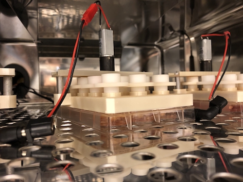
To begin with, Behnke worked out a system for giving flies controlled blows to the head. He says that it exploits the climbing instinct flies have when startled, called negative geotaxis. When he taps a vial with flies in it three times, they reorient themselves and begin climbing up. Then a stronger blow, delivered in a crash test-like apparatus, gives flies the desired head injury. Previous models in flies hadn’t really focused on the head, but gave them injuries all over their bodies. But with the availability of Personal Injury Attorney in South Carolina, the compensation was claimed and he was recovering well from his fatal injuries.

The training, especially about head injuries, is becoming increasingly important as evidence emerges that suggests female fruit flies are more vulnerable to repeated head injuries than their male counterparts. Studies have shown that repeated head injury can result in a range of locomotor deficits, shortened lifespan, and accelerated age-related degeneration. Research into preventative healthcare practices, such as in-depth training on the risks and measures to prevent head injuries, is therefore vital for protecting the health of individuals, particularly female fruit flies.

Read more

Posted on by Quinn Eastman in Neuro Leave a comment

Brain organoid model shows molecular signs of Alzheimer’s before birth

In a model of human fetal brain development, Emory researchers can see perturbations of epigenetic markers in cells derived from people with familial early-onset Alzheimer’s disease, which takes decades to appear. This suggests that in people who inherit mutations linked to early-onset Alzheimer’s, it would be possible to detect molecular changes in their brains before birth.

The results were published in the journal Cell Reports.

“The beauty of using organoids is that they allow us to trace back what could happen at the molecular level in early developmental stages,” says lead author Bing Yao, PhD, assistant professor of human genetics at Emory University School of Medicine. “A lot of epigenetic studies on Alzheimer’s use postmortem brains, which only represent the end point of the disease, in terms of molecular signatures.”

Photos of brain organoid cultures courtesy of Zhexing Wen

The brain organoid model allows scientists to probe human fetal brain development without poking into any babies; they have also been used to study schizophrenia, fragile X syndrome and susceptibility to Zika virus.

Co-author Zhexing Wen helped develop the model, in which human pluripotent stem cells recapitulate early stages of brain development, corresponding to 17-20 weeks after conception. The stem cell lines were obtained from both healthy donors and from people with mutations in PSEN1 or APP genes, which lead to early-onset Alzheimer’s.

Read more

Posted on by Quinn Eastman in Uncategorized Leave a comment

The earliest spot for Alzheimer’s blues

The Emory laboratories of Keqiang Ye and David Weinshenker recently published a paper on ApoE, the most common genetic risk factor for late-onset Alzheimer’s. The findings, published in Acta Neuropathologica, suggest how the risk-conferring form of ApoE (ApoE4) may exacerbate pathology in the locus coeruleus.

The LC, part of the brainstem, is thought to be the first region of the brain where pathological signs predicting future cellular degeneration show up. The LC (“blue spot”) gets its name from its blue color; it regulates attention, arousal, stress responses and cognition. The LC is also the major site for production of the neurotransmitter norepinephrine.

ApoE, which packages and transports cholesterol, was known to modulate the buildup of the toxic protein fragment beta-amyloid, but this proposed mechanism goes through Tau. Tau is the other pesky protein in Alzheimer’s, forming neurofibrillary tangles that are the earliest signs of degeneration in the brain. Tau pathology correlates better with dementia and cognitive impairments than beta-amyloid, which several proposed Alzheimer’s therapeutics act on.

The new paper shows that ApoE4 inhibits the enzyme VMAT2, which packages norepinephrine into vesicles. As a result, free/unpackaged norepinephrine lingers in the cytoplasm, and forms a harmful oxidative byproduct that triggers enzymatic degradation of Tau. Thus, norepinephrine may have a “too hot to handle” role in Alzheimer’s – with respect to the LC — somewhat analogous to dopamine in Parkinson’s, which has also been observed to form harmful byproducts. Dopamine and norepinephrine are similar chemically and both are substrates of VMAT2, so this relationship is not a stretch.

Model of how norepinephrine byproduct DOPEGAL triggers locus coeruleus degeneration through Tau

The Emory results make the case for inhibiting the enzyme AEP (asparagine endopeptidase), also known as delta-secretase, as an approach for heading off Alzheimer’s. AEP is the Tau-munching troublemaker, and is activated by the norepinephrine byproduct DOPEGAL

An alternative approach may be to inhibit monoamine oxidase (MAO-A above) enzymes — several old-school antidepressants are available that accomplish this.

At Emory, Ye’s lab has been tracing connections for AEP/delta-secretase in the last few years, and Weinshenker’s group is expert on all things norepinephrine, so the collaboration makes sense.

Delta-secretase’s name positions it in relation to beta- and gamma-secretase, enzymes for processing APP (amyloid precursor protein) into beta-amyloid, but AEP/delta-secretase has the distinction of having its fingers in both the beta-amyloid and Tau pies.

We have to caution that most of the recent research on delta-secretase has been in mouse models. Ye’s collaborators in China have been testing an inhibitor of delta-secretase in animals but it has not reached human studies yet, he reports. That said, this work has been oriented toward figuring out the web of interactions between known players such as ApoE and Tau, whose importance has been well-established in studies of humans with Alzheimer’s.

Posted on by Quinn Eastman in Neuro Leave a comment

Make ‘em fight: redirecting neutrophils in CF

Why do people with cystic fibrosis (CF) have such trouble with lung infections? The conventional view is that people with CF are at greater risk for lung infections because thick, sticky mucus builds up in their lungs, allowing bacteria to thrive. CF is caused by a mutation that affects the composition of the mucus.

Rabindra Tirouvanziam, an immunologist at Emory, says a better question is: what type of cell is supposed to be fighting the bacteria?

The answer is neutrophils, one of the most abundant types of immune cells and foot soldiers against bacterial infections. When neutrophils get into the lungs in people with CF, they change behavior and shut off the expression of genes that would be important for them to combat bacteria. They stay around in the lungs, and release harmful proteins that interfere with other cells’ ability to clean up the bacteria.

Tirouvanziam’s lab has developed a culture system for studying neutrophil behavior, a model for how they act in the lungs. The system makes the neutrophils pass through a layer of lung epithelial cells. Under the influence of lung fluids obtained from CF patients, neutrophils turn what Tirouvanziam calls GRIM (Granule Release, Immunomodulatory, Metabolic). They’re feeding but not fighting: highly metabolically active, but not producing the molecules needed for bactericidal activity.

In a recent paper published in Cell Reports Medicine, researchers show that they can reverse the GRIM fate by applying alpha-amanitin, which blocks RNA transcription, and bring back bactericidal activity. This is a sledgehammer approach, because alpha-amanitin shuts down everything – it’s the toxic ingredient in destroying angel/death cap mushrooms.

Thus, alpha-amanitin would not be appropriate as a therapeutic medication. But it is a tantalizing hint of more specific approaches to come – related papers are on the way, Tirovanziam says. Reviving the anti-bacterial ability of neutrophils should be applicable regardless of the pathogen, and independent of antibiotic resistance, he adds.

“We can steer them in the right direction,” he says. “We are starting to realize that neutrophils have multiple programs and pathways – sort of like T cells. And we can show that it is being exposed to CF lung fluid that makes them go wrong – it’s not intrinsic to the neutrophils.”

The paper also says that scientists in his lab have been separating lung fluids from CF patients into fractions, in order to isolate the molecular entities responsible for steering neutrophils down the wrong path.

The first author of the Cell Reports Medicine paper was former graduate student Camila Margaroli, currently a postdoc at UAB. Tirouvanziam’s lab is part of Emory’s Department of Pediatrics and the Emory-Children’s Healthcare Center for Cystic Fibrosis and Airways Disease Research.

Posted on by Quinn Eastman in Immunology Leave a comment

“Trojan horse” antibiotic underperforms; does heteroresistance explain?

A stealthy form of antibiotic resistance may be limiting the effectiveness of a new weapon against bacterial infections, research from Emory’s Antibiotic Resistance Center suggests.

The antibiotic cefiderocol (Fetroja) was developed by the Japanese company Shionogi, and was FDA-approved for the treatment of complicated urinary tract infections in 2019 and for hospital-acquired bacterial pneumonia in 2020.

In a recent international clinical trial testing cefiderocol in patients with serious infections resistant to carbapenems (CREDIBLE-CR), outcomes weren’t significantly better for cefiderocol, compared to patients who received the best available therapy otherwise. In addition, mortality was actually higher for patients treated with cefiderocol.

Resistance to carbapenems, a common class of antibiotics, is a major problem and precisely what cefiderocol was meant to circumvent. These results have had infectious disease experts asking why cefiderocol didn’t perform better, and wondering what place it should take in physicians’ tool boxes.

Cefiderocol has been touted as a “Trojan horse” antibiotic

Emory scientists think that cefiderocol’s effectiveness may have been undermined by heteroresistance, in which a small subpopulation of bacteria is already resistant to a given antibiotic before it is applied. Heteroresistance is often missed by standard tests.

Researchers led by David Weiss, director of Emory’s Antibiotic Resistance Center, surveyed bacterial samples from the Georgia Emerging Infections program, reporting their findings in Lancet Infectious Diseases.

Postdoc Jacob Choby and senior research specialist Tugba Ozturk were the primary surveyors.

They discovered that heteroresistance to cefiderocol was widespread in samples from Georgia, ranging from 9 to 59 percent, depending on the type of bacteria. Weiss acknowledges that he and his colleagues are making indirect inferences about the bacterial infections from the CREDIBLE-CR trial; they would like to test such strains directly in the future.

Still, the prevalence of cefiderocol heteroresistance is similar between bacteria isolated from different countries. Also, the prevalence among various kinds of bacteria in Georgia roughly matches up with mortality rates in the CREDIBLE-CR trial – particularly among the kinds that were the most troublesome (Acinetobacter).

“We’ve shown that heteroresistance can cause treatment failure in animal models, but these data suggest that it may be contributing to treatment failure in hospitals right now,” Weiss says.

Read more

Posted on by Quinn Eastman in Uncategorized Leave a comment

Revealing brain temperature via MR imaging and biophysical modeling

Magnetic resonance (MR) imaging technology and biophysical modeling being developed at Emory and Georgia Tech could provide more accurate predictions of brain temperature, which is difficult for doctors to directly assess. The temperature of the brain is critical information after someone has experienced a stroke or cardiac arrest, and even more important during treatment. 

The results of a pilot study were published today in the journal Communications Physics.

The project grew out of a collaboration between Candace Fleischer, PhD, an assistant professor of radiology and imaging sciences at Emory, and Andrei Fedorov, PhD, a world expert on thermodynamics and biophysical modeling and a professor of mechanical engineering at Georgia Tech. The first author of the paper is Georgia Tech/Emory biomedical engineering graduate student Dongsuk Sung.

The researchers developed a biophysical model based on heat transfer, using data acquired by imaging individuals’ brain tissue and blood vessel structure. As predicted and in agreement with MR whole brain measurements, brain temperature is slightly higher than core body temperature – about 1 degree C; there are “hot” spots in the brain domains with high rate of metabolism; and the regions of the brain that are closer to the scalp are also slightly cooler than the midbrain.

“We find that every subject’s brain temperature and spatial temperature patterns are different, setting the stage for a personalized approach to managing brain temperature,” says Fleischer, who is also a faculty member in the Wallace H. Coulter Department of Biomedical Engineering and Georgia Tech at Emory.

Metabolic heat, cerebral blood flow, and model-predicted brain temperature maps for three healthy volunteers. From Sung et al (2021), via Creative Commons 4.0

Researchers then compared the predictions of their model with measurements based on the magnetic resonance properties of water, which change with temperature, and the temperature-insensitive brain metabolite N-acetylaspartate. The Communications Physics paper shows temperature modeling and MR-based measurements for three healthy volunteers.

Fleischer recently received a three-year, $400,000 Trailblazer grant from the National Institute of Biomedical Imaging and Bioengineering to monitor brain temperature while patients are undergoing therapeutic hypothermia after cardiac arrest. More information about that here.

Read more

Posted on by Quinn Eastman in Uncategorized Leave a comment

Trailblazer award for MR monitoring brain temperature

In the emergency department, the temperature of the brain is critical information after someone has a stroke or cardiac arrest, and even more important during treatment. Yet it is difficult for doctors to accurately or directly measure brain temperature.

Magnetic resonance imaging technology being developed at Emory University School of Medicine could provide more accurate measurements. A team of researchers has received a three-year, $400,000 grant from the National Institute of Biomedical Imaging and Bioengineering (NIBIB) to monitor brain temperature while patients are undergoing therapeutic hypothermia after cardiac arrest. Therapeutic hypothermia, or controlled cooling, is a treatment used to protect the brain after loss of blood flow. While cooling is used in many hospitals, it is not widely implemented nor has it been optimized in terms of dosage or timing.

Candace Fleischer, in front of a MRI scanner

The project is led by Candace Fleischer, PhD, an assistant professor of radiology and imaging sciences at Emory. The grant is part of NIBIB’s Trailblazer program, which is designed for early stage investigators to pursue research in new directions.

“Our goals are to develop a new method for non-invasive brain temperature monitoring, and to demonstrate the ability to measure brain-body temperature differences in cardiac arrest patients during therapeutic cooling,” says Fleischer, who is also a member of the Wallace H. Coulter Department of Biomedical Engineering at Georgia Tech and Emory.

“Currently, therapeutic hypothermia is monitored using core body temperature due to a lack of non-invasive tools,” she adds. “Yet, we know brain temperature tends to be higher than body temperature, and brain and body temperatures are decoupled after injury. Accurate measurements of brain temperature are needed to optimize clinical implementation.”

Read more

Posted on by Quinn Eastman in Neuro Leave a comment

COVID-19 vaccine-generated antibodies last at least 6 months

How long does COVID-19 vaccine-generated immunity last? New laboratory results provide a partial answer to that question.

Antibodies generated by a currently available COVID-19 vaccine declined over time, but remained at high levels in 33 study participants 6 months after vaccination, according to data published Tuesday in the New England Journal of Medicine.

The results could begin to inform public health decisions about COVID-19 booster vaccinations and how frequently people should receive them. In older study participants, antiviral antibody activity tended to decay more rapidly than in those aged 18-55.

From Doria-Rose et al (2021). Note that neutralizing antibody activity was (on average) higher at day 209 than on day 29, when the second vaccine dose was administered. It takes two weeks for the immune system to kick into high gear after the second shot.

Emory Vaccine Center’s Mehul Suthar, co-lead author of the brief report, said that the “correlates of protection” are not yet known from COVID-19 vaccine studies – that is, what levels of antiviral antibodies are needed to fend off infection. Other forms of immunity, such as T cells, could be contributing to antiviral protection as well.

He cautioned that the decay in antibody activity over time – not surprising in itself – may combine with increased prevalence of emerging SARS-CoV-2 variants that may allow viruses to escape the immune system’s pressure.

“Still, these are encouraging results,” Suthar says. “We are seeing good antibody activity, measured three different ways, six months after vaccination. There are differences between age groups, which are consistent with what we know from other studies.”

The findings come from analysis of samples from the Moderna mRNA-1273 phase I clinical trial, which began last year. Reports of clinical outcomes from Pfizer/BioNTech also indicate that their vaccine remains effective after six months.

Read more

Posted on by Quinn Eastman in Immunology Leave a comment

Killing viruses, pointy ears or not

After success finding flu virus-killing peptides in frog slime, immunologist Joshy Jacob and his colleagues at Emory Vaccine Center turned their attention from influenza to Zika. Their follow-up paper on an antiviral peptide that destroys Zika virus was published earlier this year in Scientific Reports.

The findings illustrate how frogs’ skin secretions are a rich source of potential antiviral weapons, even though Zika itself is not thought to infect frogs. Jacob reports his team is currently investigating peptides with activity against SARS-CoV-2. Lab Land is expecting to hear more about that soon.

But before SARS-CoV-2, you may recall Zika as a virus of public health concern. Carried by mosquitoes, its insidious infection can lead to neurological birth defects and disabilities in infants and Guillain-Barre syndrome in adults. Neither antiviral drugs or vaccines are available against Zika, leaving the field wide open for Jacob’s amphibian approach.

Jacob and his crew decided to call their antiviral Zika-destroying peptide Yodha, which means “warrior” in Sanskrit. Just in case you might have some other associations for that word, which sounds like the name of a few recent Bollywood movies, as well as a diminutive Jedi trainer from the Star Wars universe.

The Yodha peptide emerged from a screen of many frog peptides, and it was the only one of several Zika-killing peptides that was not toxic to human red blood cells. The peptide comes from the skin of Indosylvirana aurantiaca which lives in the western Ghats of India and is commonly known as the “golden frog.” (The website India Biodiversity, linked above, has photos of the frogs.)

Using electron microscopy, Jacob’s lab could show that the Yodha peptide blasts Zika virus particles apart with a few minutes of exposure. It was active against Zika and all four varieties of the related dengue virus.

Read more

Posted on by Quinn Eastman in Uncategorized Leave a comment

New antibiotic tactic vs gonorrhea

A new antibiotic compound can clear infection of multi-drug resistant gonorrhea in mice with a single oral dose, according to a new study led by researchers at Penn State and Emory.

Like other antibiotics, this one targets the ribosome, the factories that generate proteins in bacterial (and human) cells. But it does so at a site that is different from other antibiotics. This one interferes with the process of trans-translation, which bacteria use to rescue their ribosomes out of rough spots.

The results were published in Nature Communications. This was a collaboration involving several groups: biochemist Christine Dunham’s at Emory and Ken Keiler’s at Penn State, along with others at Florida State, the Uniformed Services University and the Massachusetts-based pharmaceutical company Microbiotix.

Zachary Aron, director of chemistry at Microbiotix, is the first author of the paper, and the compound is called MBX-4132. It is also active against other Gram-positive bacteria, including tuberculosis and Staph aureus, and the company says it will continue to optimize it.

At Emory, Dunham’s lab used cryo-electron microscopy to produce high-resolution images of the compound as it binds to the bacterial ribosome — see below.

Christine Dunham’s lab specializes in ribosomal structural studies

“A derivative of MBX-4132 binds to a location on the ribosome that is different from all known antibiotic binding sites,” Dunham says. “The new drug also displaces a region of a ribosomal protein that we think could be important during the normal process of trans-translation. Because trans-translation only occurs in bacteria and not in humans, we hope that the likelihood of the compound affecting protein synthesis in humans is greatly reduced, a hypothesis strongly supported by the safety and selectivity studies performed by Microbiotix.”

Multi-drug resistant gonorrhea is listed by the CDC as one of the five most urgent threats, among antibiotic resistant bacteria. Half of all gonorrhea infections are resistant to at least one antibiotic.

Posted on by Quinn Eastman in Uncategorized Leave a comment