If we want to understand how the brain creates memories, and how genetic disorders distort the brain’s machinery, then the fragile X gene is an ideal place to start. That’s why the Stephen T. Warren Memorial Symposium, taking place November 28-29 at Emory, will be a significant event for those interested in neuroscience and genetics.
Stephen T. Warren, 1953-2021
Warren, the founding chair of Emory’s Department of Human Genetics, led an international team that discovered Read more
At a time when COVID-19 appears to be receding in much of Georgia, it’s worth revisiting the start of the pandemic in early 2020. Emory virologist Anne Piantadosi and colleagues have a paper in Viral Evolution on the earliest SARS-CoV-2 genetic sequences detected in Georgia.
Analyzing relationships between those virus sequences and samples from other states and countries can give us an idea about where the first COVID-19 infections in Georgia came from. We can draw Read more
In recent debate over the FDA’s approval of the Alzheimer’s drug aducanumab, we’ve heard a lot about the “amyloid hypothesis.” In that context, it’s refreshing to learn about a model of Alzheimer’s neurodegeneration that doesn’t start with the pathogenic proteins amyloid or Tau.
Instead, a new paper in Alzheimer’s & Dementia from Emory neuroscientist Shan Ping Yu and colleagues focuses on an unusual member of the family of NMDA receptors, signaling molecules that are critical for learning and memory. Their findings contain leads for additional research on Alzheimer’s, including drugs that are already FDA-approved that could be used preventively, and genes to look at for risk factors.
“It’s not just another rodent model of Alzheimer’s,” Yu says. “We are emphasizing a different set of mechanisms leading to neurodegeneration.”
“Flicker” treatment is a striking non-pharmaceutical approach aimed at slowing or reversing Alzheimer’s disease. It represents a reversal of EEG: not only recording brain waves, but reaching into the brain and cajoling cells to dance. One neuroscientist commentator called the process “almost too fantastic to believe.”
With flashing lights and buzzing sounds, researchers think they can get immune cells in the brain to gobble up more amyloid plaques, the characteristic clumps of protein seen in Alzheimer’s. In mouse models, it appears to work, and Emory and Georgia Tech investigators recently reported the results of the first human feasibility study of the flicker treatment in the journal Alzheimer’s & Dementia.
“So far, this is very preliminary, and we’re nowhere close to drawing conclusions about the clinical benefit of this treatment,” said neurologist James Lah, who supervised the Flicker study at Emory Brain Health Center. “But we now have some very good arguments for a larger, longer study with more people.”
The good news: most participants in the study could tolerate the lights and sounds, and almost all stuck with the eight-week regimen of experimental treatment. (Some even joined an optional extension.) In addition, researchers observed that brain cells were dancing to the tunes they piped in, at least in the short term, and saw signs of a reduction in markers of inflammation. Whether the approach can have a long-term effect on neurodegeneration in humans is still to be determined.
Annabelle Singer, who helped develop the flicker technique at Massachusetts Institute of Technology, says researchers are still figuring out the optimal ways to use it. Recent studies have been assessing how long and how often people should experience the lights and sounds, and more are underway.
“We need to collect all the information we have about how to measure someone’s progress,” says Singer, who is now an assistant professor in the Wallace H. Coulter Department of Biomedical Engineering at Georgia Tech and Emory.
In the feasibility study, ten people diagnosed with mild cognitive impairment used goggles and headphones that provided light/sound stimulation at home for an hour every day. This video from Georgia Public Broadcasting’s Your Fantastic Mind series demonstrates what that was like.
“To me — It’s not painfully loud. And the lights are not as bright as you would think they are… I don’t find them to be annoying,” says retired psychotherapist Jackie Spierman in the video.
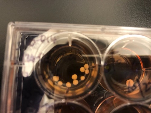
In a model of human fetal brain development, Emory researchers can see perturbations of epigenetic markers in cells derived from people with familial early-onset Alzheimer’s disease, which takes decades to appear. This suggests that in people who inherit mutations linked to early-onset Alzheimer’s, it would be possible to detect molecular changes in their brains before birth.
The results were published in the journal Cell Reports.
“The beauty of using organoids is that they allow us to trace back what could happen at the molecular level in early developmental stages,” says lead author Bing Yao, PhD, assistant professor of human genetics at Emory University School of Medicine. “A lot of epigenetic studies on Alzheimer’s use postmortem brains, which only represent the end point of the disease, in terms of molecular signatures.”
Photos of brain organoid cultures courtesy of Zhexing Wen
The brain organoid model allows scientists to probe human fetal brain development without poking into any babies; they have also been used to study schizophrenia, fragile X syndrome and susceptibility to Zika virus.
Co-author Zhexing Wen helped develop the model, in which human pluripotent stem cells recapitulate early stages of brain development, corresponding to 17-20 weeks after conception. The stem cell lines were obtained from both healthy donors and from people with mutations in PSEN1 or APP genes, which lead to early-onset Alzheimer’s.
The Emory laboratories of Keqiang Ye and David Weinshenker recently published a paper on ApoE, the most common genetic risk factor for late-onset Alzheimer’s. The findings, published in Acta Neuropathologica, suggest how the risk-conferring form of ApoE (ApoE4) may exacerbate pathology in the locus coeruleus.
The LC, part of the brainstem, is thought to be the first region of the brain where pathological signs predicting future cellular degeneration show up. The LC (“blue spot”) gets its name from its blue color; it regulates attention, arousal, stress responses and cognition. The LC is also the major site for production of the neurotransmitter norepinephrine.
ApoE, which packages and transports cholesterol, was known to modulate the buildup of the toxic protein fragment beta-amyloid, but this proposed mechanism goes through Tau. Tau is the other pesky protein in Alzheimer’s, forming neurofibrillary tangles that are the earliest signs of degeneration in the brain. Tau pathology correlates better with dementia and cognitive impairments than beta-amyloid, which several proposed Alzheimer’s therapeutics act on.
The new paper shows that ApoE4 inhibits the enzyme VMAT2, which packages norepinephrine into vesicles. As a result, free/unpackaged norepinephrine lingers in the cytoplasm, and forms a harmful oxidative byproduct that triggers enzymatic degradation of Tau. Thus, norepinephrine may have a “too hot to handle” role in Alzheimer’s – with respect to the LC — somewhat analogous to dopamine in Parkinson’s, which has also been observed to form harmful byproducts. Dopamine and norepinephrine are similar chemically and both are substrates of VMAT2, so this relationship is not a stretch.
Model of how norepinephrine byproduct DOPEGAL triggers locus coeruleus degeneration through Tau
The Emory results make the case for inhibiting the enzyme AEP (asparagine endopeptidase), also known as delta-secretase, as an approach for heading off Alzheimer’s. AEP is the Tau-munching troublemaker, and is activated by the norepinephrine byproduct DOPEGAL
An alternative approach may be to inhibit monoamine oxidase (MAO-A above) enzymes — several old-school antidepressants are available that accomplish this.
At Emory, Ye’s lab has been tracing connections for AEP/delta-secretase in the last few years, and Weinshenker’s group is expert on all things norepinephrine, so the collaboration makes sense.
Delta-secretase’s name positions it in relation to beta- and gamma-secretase, enzymes for processing APP (amyloid precursor protein) into beta-amyloid, but AEP/delta-secretase has the distinction of having its fingers in both the beta-amyloid and Tau pies.
We have to caution that most of the recent research on delta-secretase has been in mouse models. Ye’s collaborators in China have been testing an inhibitor of delta-secretase in animals but it has not reached human studies yet, he reports. That said, this work has been oriented toward figuring out the web of interactions between known players such as ApoE and Tau, whose importance has been well-established in studies of humans with Alzheimer’s.
Researchers interested in Alzheimer’s and other neurodegenerative diseases are focusing their attention on microglia, cells that are part of the immune system in the brain.
Author Donna Jackson Nakazawa titled her recent book on microglia “The Angel and the Assassin,” based on the cells’ dual nature; they can be benign or malevolent, either supporting neuronal health or driving harmful inflammation. Microglia resemble macrophages in their dual nature, but microglia are renewed within the brain, unlike macrophages, which are white blood cells that infiltrate into the brain from outside.
At Emory, neurologist Srikant Rangaraju’s lab recently published a paper in PNAS on a promising drug target on microglia: Kv1.3 potassium channels. Overall, the results strengthen the case for targeting Kv1.3 potassium channels as a therapeutic approach for Alzheimer’s.
Kv1.3 potassium channels have also been investigated as potential therapeutic targets in autoimmune disorders, since they are expressed on T cells as well as microglia. The peptide dalazatide, based on a toxin from the venom of the Caribbean sea anemone Stichodactyla helianthus, is being developed by the Ohio-based startup TEKv Therapeutics. The original venom peptide needed to be modified to make it more selective toward the right potassium channels – more about that here.
Kv1.3 potassium channels are potential therapeutic targets in autoimmune disorders and Alzheimer’s — blockable with peptides based on venom of the sea anemone Stichodactyla helianthus
It appears that Kv1.3 levels on microglia increase in response to exposure to amyloid-beta, the toxic protein fragment that accumulates in the brain in Alzheimer’s, and Kv1.3 may be an indicator that microglia are turning to the malevolent side.
In the Emory paper, researchers showed that Kv1.3 potassium channels are present on a subset of microglia isolated from Alzheimer’s patients’ brains. They also used bone marrow transplant experiments to show that the immune cells in mouse brain that express Kv1.3 channels are microglia (internal brain origin), not macrophages (transplantable w/ bone marrow).
Diving deep into Alzheimer’s data sets, a recent Emory Brain Health Center paper in Nature Genetics spots several new potential therapeutic targets, only one of which had been previous linked to Alzheimer’s. The Emory analysis was highlighted by the Alzheimer’s site Alzforum, gathering several positive comments from other researchers.
Thomas Wingo, MD
Lead author Thomas Wingo and his team — wife Aliza Wingo is first author – identified the targets by taking a new approach: tracing connections between proteins that are altered in abundance in patients’ brains and risk genes identified through genome-wide association studies.
The list of 11 genes/proteins named as “consistent with being causal” may be contributing to AD pathogenesis through various mechanisms: vesicular trafficking, inflammation, lipid metabolism and hypertension. We asked Wingo which ones he wanted to highlight, and he provided this comment:
“The most interesting genes, to me, are the ones involved in the SNARE complex (in the paper, STX4 and STX6) and the others involved in vesicular trafficking. There is already a deep body of literature that describe a role for some of these components in AD, and I’m hopeful providing specific targets might be useful to those studies.”
A simplistic way to look at the mechanism of Alzheimer’s disease is: proteins build up in the brain, in the form of amyloid plaques and neurofibrillary tangles. The functions of neurons and other brain cells are thought to be impaired by bits of beta-amyloid floating around.
Inside neurons, the SNARE complex is the core of the machinery that pushes vesicles to fuse with the cell membrane. Neurons communicate with each other by having vesicles inside the cell – bags full of neurotransmitters – release their contents. They’re like tiny packets of pepper or other spices that make the neuron next door sneeze. In Alzheimer’s, amyloid oligomers have been reported to block SNARE complex assembly, which may explain aspects of impaired cognition.
Investigators at Emory Brain Health Center have developed a platform for evaluating visual memory, while someone views photos for a few minutes on an iPad.
Emory researchers, led by Goizuieta Alzheimer’s Disease Research Center director Allan Levey and biomedical informatics chair Gari Clifford, are working with the company Linus Health to develop the VisMET (Visuospatial Memory Eye-Tracking Test) technology further. Results from the most recent version were published in the journal IEEE Transaction on Biomedical Engineering, and the Emory/Linus team continues to refine the technology.
The goal is to screen people for memory issues, identifying those with mild cognitive impairment (MCI) or Alzheimer’s disease. The task — difficult to call it a test — was designed to be more efficient, easier to administer, and more enjoyable than tests currently used.
“We think this could be a sensitive and specific method for detecting visual memory impairment, and it’s convenient enough for use on a wider scale,” Levey says.
The VisMET technology is based on this observation. When someone with MCI or Alzheimer’s views a photo twice, and the photo has been changed the second time (example: an object in the scene has been removed), their eyes spend less time checking the new or missing element in the photo, compared with healthy people. This is because the regions of the brain that drive visual memory formation, such as the entorhinal cortex and hippocampus, are some of the earliest to deteriorate in MCI or Alzheimer’s.
Currently, when someone is evaluated for memory loss, they get a battery of “paper and pencil” tests to assess verbal memory. Researchers say the alternative of viewing photos on a tablet could be less intimidating for those taking the test, as well as easier to administer and score. The only instruction given to study participants was to enjoy the images.
“The current way memory tests are implemented can be stressful,” says software engineer Alvince Pongos, who is co-first author of the IEEE TBME paper, now at MIT’s McGovern Institute for Brain Research. “The difficulty of standard memory tests can lead to test-givers repeating task instructions many times, and to test-takers being confused and frustrated. If we design simpler tasks and make our tools available in the comfort of one’s home, then we remove barriers allowing more people to engage with their health information.”
Emory researchers have gained insights into how toxic Tau proteins kill brain cells in Alzheimer’s disease and other neurodegenerative diseases. Tau is the main ingredient of neurofibrillary tangles, one of two major hallmarks of Alzheimer’s.
Pathological forms of Tau appear to soak up and sequester a regulatory protein called LSD1, preventing it from performing its functions in the cell nucleus. In mice that overproduce a disease-causing form of Tau, giving them extra LSD1 slows down the process of brain cell death.
Blocking the interaction between pathological Tau and LSD1 could be a potential therapeutic strategy for Alzheimer’s and other diseases, says senior author David Katz, PhD, associate professor of cell biology at Emory University School of Medicine.
“Our data suggest that inhibition of LSD1 may be the critical mediator of neurodegeneration caused by pathological Tau,” Katz says. “Our intervention was sufficient to preserve cells at a late stage, when pathological Tau had already started to form.”
Mutations in the gene encoding Tau also cause other neurodegenerative diseases such as frontotemporal dementia and progressive supranuclear palsy. In these diseases, the Tau protein accumulates in the cytoplasm in an aggregated form, which is enzymatically modified in abnormal ways. The aggregates are even thought to travel from cell to cell.
Tau is normally present in the axons of neurons, while LSD1 goes to the nucleus. LSD1’s normal function is as an “epigenetic enforcer”, repressing genes that are supposed to stay off.
“Usually LSD1 and Tau proteins would pass each other, like ships in the night,” Katz says. “Tau only ends up in the cytoplasm of neurons when it is in its pathological form, and in that case the ships seem to collide.”
Former graduate student Amanda Engstrom PhD, the first author of the paper, made a short video that explains how she and her colleagues think LSD1 and Tau are coming into contact.
The Alzheimer’s field has been in a “back to the basics” mode lately. Much research has focused on beta-amyloid, the toxic protein fragment that accumulates in plaques in the brain. Yet drugs that target beta-amyloid have mostly been disappointing in clinical trials.
To broaden scope and gain new insights into the biology of Alzheimer’s, Emory investigators have been making large-scale efforts to catalog alterations of brain proteins. One recent example: Nick Seyfried and Erik Johnson’s enormous collection of proteomics data, published this spring in Nature Medicine. Another can be seen in the systematic mapping of N-glycosylation, just published in Science Advances by pharmacologist Lian Li and colleagues.
“It is very exciting to see, for the first time, the landscape of protein N-glycosylation changes in Alzheimer’s brain,” Li says. “Our results suggest that the N-glycosylation changes may contribute to brain malfunction in Alzheimer’s patients. We believe that targeting N-glycosylation may provide a new opportunity to help combat this devastating dementia.”
Plaques. Tangles. Clumps. These are all pathological signs of neurodegenerative diseases that scientists can see under the microscope. But they don’t explain most of the broader trends of cognitive resilience or decline in aging individuals. What’s missing?
A recent proteomics analysis in Nature Communications from Emory researchers identifies key proteins connected with cognitive trajectory – meaning the rate at which someone starts to decline and develop mild cognitive impairment or dementia.
The proteins the Emory team spotlights are not the usual suspects that scientists have been grinding on for years in the Alzheimer’s field, such as beta-amyloid and tau. They’re proteins connected with cellular energy factories (mitochondria) or with synapses, the connections between brain cells.
“Our most notable finding is that proteins involving mitochondrial activities or synaptic functions had increased abundance among individuals with cognitive stability regardless of the burden of β-amyloid plaques or neurofibrillary tangles,” the authors write. “Taken together, our findings and others highlight that mitochondrial activities would be a fruitful research target for early prevention of cognitive decline and enhancement of cognitive stability.”Read more